ECR 2017 / C-2435
High-frequency ultrasound of the ankle.
Congress:
ECR 2017
Poster Number:
C-2435
Type:
Educational Exhibit
Keywords:
Anatomy, Musculoskeletal system, Ultrasound, Education, Structured reporting, Education and training
Authors:
C. M. Olchowy, D. Soliński, A. Niemczyk, K. Niemczyk, A. Zwoliński , K. Cebulski, A. Olchowy, M. Łasecki, U. Zaleska-Dorobisz; Wroclaw/PL
DOI:
10.1594/ecr2017/C-2435
")
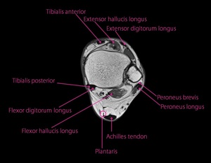
Fig. 1:
Tendons of the ankle joint. Axial MRI scan.
")
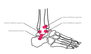
Fig. 2:
Lateral ankle ligaments.
")
Fig. 3:
4 compartments of the ankle. Structures visible in Ultrasound imaging.
")
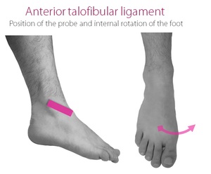
Fig. 4:
Anterior talofibular ligament. Position of the probe. Internal deviation of the...
")
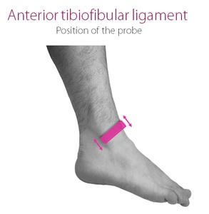
Fig. 5:
Anterior tibiofibular ligament.
Position of the probe.
")
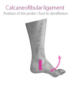
Fig. 6:
Calcaneofibular ligament. The probe position. Tendon is best seen in...
")
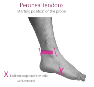
Fig. 7:
Peroneal tendons . Short-axis view on the level of the lateral malleolus....
")
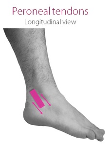
Fig. 8:
Peroneal tendons. Probe position. Long-axis view on the level of lateral...
")
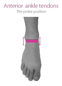
Fig. 9:
Anterior Ankle Tendons. Probe position.
")
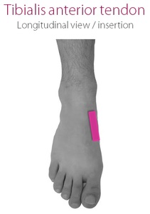
Fig. 10:
Probe positioning in evaluation of distal part of Tibialis anterior tendon and...
")
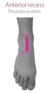
Fig. 11:
Anterior tibio-talar recess - the probe position.
")
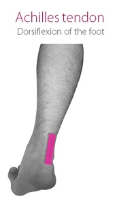
Fig. 12:
Achilles tendon. Longitudinal view.
")
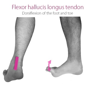
Fig. 13:
Flexor hallucis longus probe positioning. Please note, that foot and tirst toe...
")
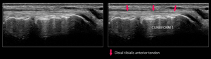
Fig. 15:
Distal tibialis tendon and its insertion.
")
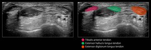
Fig. 16:
Anterior ankle tendons - transverse view
")
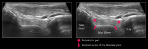
Fig. 17:
Anterior tibio-talar recess.
")
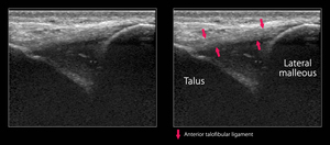
Fig. 18:
Anterior talofibular ligament. The most frequently injured ligament of the...
")
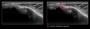
Fig. 19:
Anterior tibiofibular ligament.
")
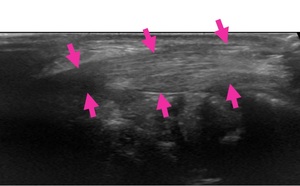
Fig. 20:
Peroneus longus and peroneus brevis tendons. Long-axis scan.
")
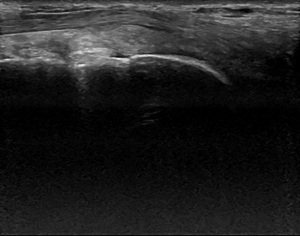
Fig. 21:
Flexor hallucis longus tendon.
")
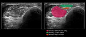
Fig. 22:
Peroneus longus and peroneus brevis tendons on the level of lateral maleolus....
")
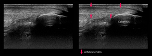
Fig. 23:
Achilles tendon. Long-axis scan.
")
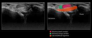
Fig. 24:
Calcaneofibular ligament. Please note, that peroneal tendons are superficial to...