Type:
Educational Exhibit
Keywords:
Pathology, Neoplasia, Diagnostic procedure, Education, MR, CT, Pancreas, Biliary Tract / Gallbladder, Abdomen, MR-Cholangiography
Authors:
J. Munechika1, N. Mizobuchi1, Y. Ohgiya1, T. Gokan1, N. Takeyama2, E. Tanaka2, N. Ohike2, T. Norose2; 1Tokyo/JP, 2Yokohama/JP
DOI:
10.1594/ecr2018/C-0803
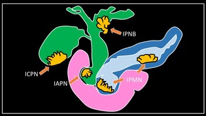
Background
In the WHO 2010 classification of tumors,
macroscopically recognizable papillary neoplastic lesions in the biliary system,
as counterparts of pancreatic intraductal papillary mucinous neoplasms (IPMN),
were described: lesions in the bile duct were defined as intraductal papillary neoplasm of bile duct (IPNB) and those in the gallbladder as intracholecystic papillary tubular neoplasm (ICPN).
Papillary neoplastic lesions in the papilla of Vater,
proliferating in the papillary duct (common channel–intrapapillary bile duct/pancreatic duct) and differentiating from the surrounding mucosa,
have also been proposed and are collectively referred to as intraampullary papillary tubular neoplasm (IAPN).
These neoplasms are regarded as precancerous or preinvasive cancerous lesions,
and distinguished from conventional invasive or papillary adenocarcinomas.
Fig. 1: Classification of intraductal papillary neoplasms of pancreas, bile duct, gallbladder, and ampulla