Keywords:
Ear / Nose / Throat, Paediatric, Head and neck, CT-High Resolution, CT, Statistics, Congenital, Dysplasias
Authors:
M. Almuntashri; Riyadh, Riyadh/SA
DOI:
10.1594/ecr2018/C-1473
Methods and materials
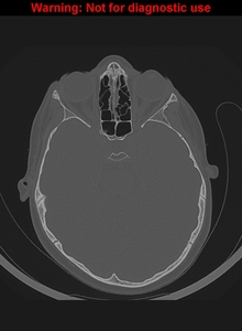
Fig. 3: Fig 3: Evaluation of the pinna on axial image.
References: Dr. Makki Almuntashri
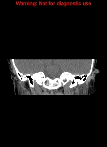
Fig. 2: Fig 2: Coronal soft tissue window of HRCT for same infant confirming the shape and size of small left pinna.
References: Dr. Makki Almuntashri
Fig. 2: Fig 2: Coronal soft tissue window of HRCT for same infant confirming the shape and size of small left pinna.
References: Dr. Makki Almuntashri
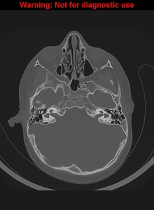
Fig. 1: This is HRCT image of 0 month old infant with bilateral microtia (grade 3) associated with atresia of external auditory canal, hypoplastic fused ossicles and deformed vestibules bilaterally.
References: Dr. Makki Almuntashri
Study design: we retrospectively included all patients with microtia who had CT scan of temporal bone from 2009 to 2016.
Source: we used PACS of medical imaging department in KAUH.
Expert certified Radiologist reviewed all patients and determined the findings on CT scan.
Data collection: all data collected on excel sheet and saved on password secured computer in the medical imaging department.
The statistician analyzed the findings.
