Type:
Educational Exhibit
Keywords:
Trauma, Education and training, Education, Ultrasound, MR, Musculoskeletal joint, Extremities, Anatomy
Authors:
R. M. L. S. Roose, A. L. A. Milants, N. Moyson, S. Provyn, M. De Maeseneer; Brussels/BE
DOI:
10.1594/ecr2018/C-2245
Background
The hallux supports 50 % of the body weight,
which can increase eightfold with jumping.
Unlike the hand in which supination is possible,
the foot is always in pronation.
Remarkably,
very similar muscles are present in the foot as in the thenar of the hand,
although their function in the foot is only flexion and extension.
The great toe is adducted and has no grasping function.
In many anatomy textbooks the anatomy of the hallux and its metatarsophalangeal joint is not described into detail.
The anatomy is complex and is challenging to adequately image.
The anatomy of the big toe is also quite different from that of the lesser toes.
The plantar plate of the smaller toes consists of a meniscoid cartilaginous structure in the midline,
whereas the ‘plantar plate’ of the big toe is made up of multiple ligaments and tendons,
and of interest,
abscent of a strong midline meniscoid structure.
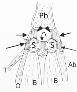
Fig. 9: Drawing of plantar view of the hallux structures. The flexor hallucis brevis (B) inserts on to the sesamoids. Medially the abductor tendon is adjacent to the medial belly of the flexor hallucis brevis (Ab). Laterally the adductor tendon is adjacent to the lateral belly of the flexor hallucis brevis belly (O, oblique head; T, transverse head). In between the sesamoids (S) the intersesamoid ligament (curved arrow) is seen. The sesamoids are connected to the phalanx (Ph) by the sesamophalangeal ligaments (arrowheads). Alongside fibers of the abductor and adductor can be seen (short arrows). The cut sesamometatarsal ligaments (long arrows) extend from the sesamoids to the metatarsals.
References: Annemieke Milants, Md. UZ Brussel-ASZ Aalst/BE

 inserts on to the sesamoids. Medially the abductor tendon is adjacent to the medial belly of the flexor hallucis brevis (Ab). Laterally the adductor tendon is adjacent to the lateral belly of the flexor hallucis brevis belly (O, oblique head; T, transverse head). In between the sesamoids (S) the intersesamoid ligament (curved arrow) is seen. The sesamoids are connected to the phalanx (Ph) by the sesamophalangeal ligaments (arrowheads). Alongside fibers of the abductor and adductor can be seen (short arrows). The cut sesamometatarsal ligaments (long arrows) extend from the sesamoids to the metatarsals. References: Annemieke Milants, Md. UZ Brussel-ASZ Aalst/BE")