ECR 2018 / C-2260

All roads lead to R..ight atrium: general review of congenital anomalies of the systemic venous return
Congress:
ECR 2018
Poster Number:
C-2260
Type:
Educational Exhibit
Keywords:
Education and training, Normal variants, MR-Angiography, CT-Angiography, CT, Veins / Vena cava, Cardiovascular system, Anatomy
Authors:
J. D. Oliveira1, I. Martins2; 1Amadora/PT, 2Lisboa/PT
DOI:
10.1594/ecr2018/C-2260

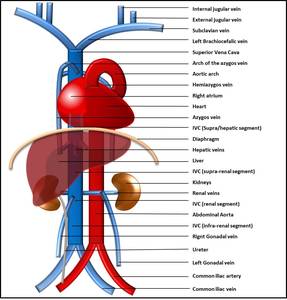
Fig. 1:
Normal systemic venous system schematic.

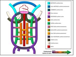
Fig. 2:
Systemic venous system development composite schematic.

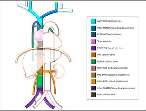
Fig. 3:
Normal sistemic venous system embryology.