ECR 2018 / C-2418
Decoding the Contrast Enhanced Brain
Congress:
ECR 2018
Poster Number:
C-2418
Type:
Educational Exhibit
Keywords:
Neoplasia, Infection, Education and training, Education, Contrast agent-intravenous, MR, Neuroradiology brain, Contrast agents
Authors:
V. B. Pai, K. Joshi, K. Gupta, C. Trivedi, A. Agrawal, B. Pai, R. Kharche, D. Lokhande; Mumbai/IN
DOI:
10.1594/ecr2018/C-2418

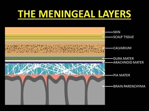
Fig. 1:
THE MENINGEAL LAYERS

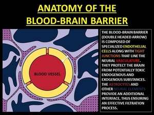
Fig. 2:
ANATOMY OF THE BLOOD-BRAIN BARRIER

Fig. 3:
PHYSIOLOGY OF THE BLOOD-BRAIN BARRIER

Fig. 4:
PHYSICS OF T1 RELAXATION AND GADOLINIUM EFFECTS ON T1 RELAXATION

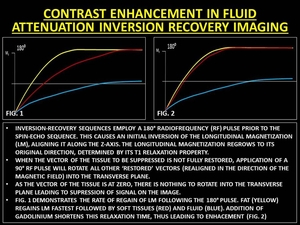
Fig. 5:
PHYSICS OF FLAIR IMAGING AND CONTRAST ENHANCEMENT IN FLAIR IMAGING