ECR 2018 / C-2529
Pictorial assay of MRI imaging findings in Pott's Spine
Congress:
ECR 2018
Poster Number:
C-2529
Type:
Scientific Exhibit
Keywords:
Infection, Education, MR, Musculoskeletal spine
Authors:
K. Tyagi, M. Varunya, A. Balachandran, S. Ashwathappa, R. HV; Bangalore/IN
DOI:
10.1594/ecr2018/C-2529

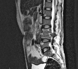
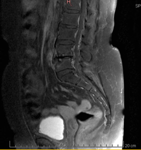
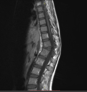
Fig. 1:
MRI image of Lumbar spine

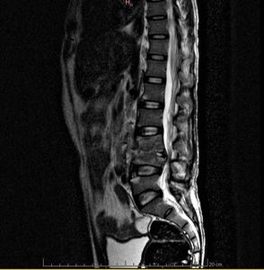
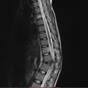
Fig. 2:
T2 WEIGHTED SAGITTAL SECTION OF SPINE

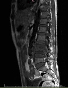
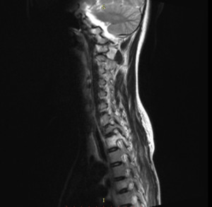
Fig. 3:
T1 WEIGHTED SAGITTAL SECTION OF SPINE

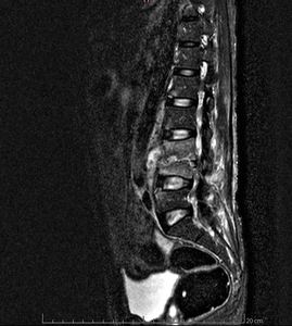
Fig. 4:
STIR SEQUENCE OF SPINE,SAGITTAL SECTION

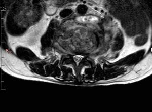
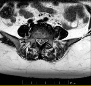
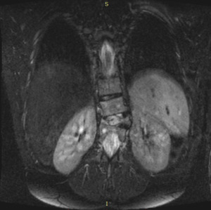
Fig. 5:
AXIAL SECTION OF SPINE T2 WEIGHTED SEQUENCE

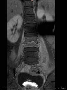
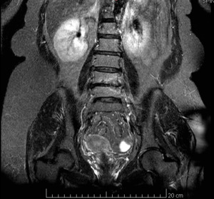
Fig. 6:
POST CONTRAST CORONAL VIEW OF SPINE

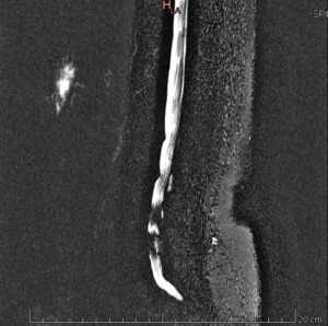
Fig. 7:
MYELOGRAM OF SPINE SAGITTAL VIEW

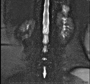
Fig. 8:
MYLEOGRAM OF SPINE CORONAL VIEW

Fig. 9:
POST CONTRAST SAGITTAL VIEW OF SPINE

Fig. 10:
STIR SEQUENCE OF SPINE

Fig. 11:
T2 WEIGHTED AXIAL SECTION OF SPINE

Fig. 12:
T2 WEIGHTED SEQUENCE OF SPINE SAGITTAL VIEW

Fig. 13:
TI WEIGHTED SEQUENCE OF SPINE SAGITTAL VIEW

Fig. 14

Fig. 19