In this section we will describe the functional anatomy of the facial nerve as well as the anatomic landmarks that will aid the general radiologist to identify it and separate normal findings from disorders.
The facial nerve (cranial nerve-CN VII) is a mixed nerve with motor,
sensory and parasympathetic fibers.
Motor branches: innervation of the
- occipitalis muscle
- posterior auricular muscle
- stylohyoid muscle
- posterior belly of the digastric muscle
- stapedius muscle
- muscles of facial expression
Sensory branches:
- sensation of taste from the anterior two-thirds of the tongue
- afferent innervation to the oropharynx below the palatine tonsil
- sensation from the ear canal,
the auricle and the retro-auricular region
Parasympathetic branches: innervation of the
- submandibular and sublingual glands
- lacrimal glands
- mucosal membranes of the nasal cavity and palate
The path of the facial nerve (Figure 1)
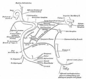
Fig. 1: Plan of the facial nerve.
References: Henry Vandyke Carter - Henry Gray (1918) Anatomy of the Human Body. Gray's Anatomy, Plate 788
The intra-axial segment (Figure 2)
It is located within the brainstem and consists of the motor nucleus (the motor fibres of the facial nerve loop posteriorly over the abducens nerve nucleus and they form the facial colliculus in the floorof fourth ventricle),
superiorsalivary nucleus (parasympathetic),
and nucleus of tractus solitarius (sensory).
These nuclei are not radiologically distinguishable from the rest of the brainstem on Computed Tomography (CT) or on state-of-the-art clinical Magnetic Resonance Imaging (MRI).
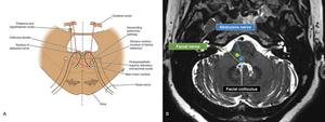
Fig. 2: The intra-axial segment. (A) Illustration of the facial nerve nuclei. (B) MRI axial T2 showing the loop of the motor fibres of the facial nerve posteriorly over the abducens nerve.
References: Richard S. Snell Clinical Neuroanatomy (7th Ed.), Radiopaedia (Case courtesy of A.Prof Frank Gaillard, Radiopaedia.org, rID: 50811),
The cisternal segment (brainstem to internal auditory canal).
(Figure 3)
Both the motor root of the facial nerve and the nervus intermedius (which contains the parasympathetic root and special sensory root of the facial nerve) leave the brainstem near the dorsal pons at the pontomedullary junction.
Within the cerebellopontine angle,
the nerve travels anterolaterally into the porus acusticus of the internal auditory canal (IAC),
anterior to the vestibulocochlear nerve.
This segment is 24mm.
The cisternal segment has no branches.
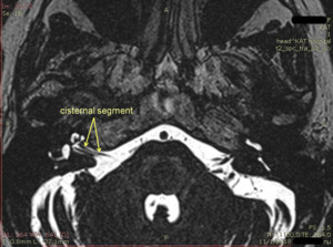
Fig. 3: The cisternal segment.
References: Department of Radiology, KAT General Hospital, Athens, Greece
The meatal (canalicular) segment (within the IAC).
(Figure 4)
This portion of the facial nerve still comprises the nervus intermedius and the facial nerve proper in this segment (a division that is often,
though not always,
poorly characterized radiologically) as it enters the IAC alongside the subcomponents of CN VIII,
the cochlear and superior and inferior vestibular nerves.
The facial nerve is consistently oriented anterior to the superior and inferior vestibular nerves and superior to the cochlear nerve.
To best evaluate the presence of all four of these nerves,
typically a high-resolution T2-weighted thin-section MRI in the sagittal plane with a cross-sectional view of the IAC is necessary as axial views show at most only two of the four nerves in a single axial plane.
The meatal segment has no branches.
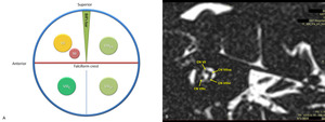
Fig. 4: Orientation of the meatal facial nerve. (A) The diagram for the orientation of the nerves of the internal acoustic meatus. (B) The annotated sagitally T2-weighted, thin-slice MR image of the internal auditory canal showing the facial nerve situated at the anterior superior quadrant of the internal auditory canal. (VII = facial nerve, NI = nervus intermedius, VIIIc = cochlear nerve, VIIIvs = superior division of vestibular nerve, VIIIvi = inferior division of vestibular nerve)
References: Radiopaedia (Case courtesy of A.Prof Frank Gaillard, Radiopaedia.org, rID: 36049), Department of Radiology, KAT General Hospital, Athens, Greece
The labyrinthine segment (IAC to geniculate ganglion).
(Figure 5)
It is both the narrowest (<0.7mm diameter) and shortest (3–5mm length) segment.
At the end of the IAC,
the facial nerve enters the facial (Fallopian) canal (bony canal from the IAC to the stylomastoid foramen)(Figure 6) on the anterior aspect of the Bill bar (tiny triangular bone separating the facial nerve from the superior vestibular nerve).
The labyrinthine segment courses superior to the cochlea and anterior to the vestibule and then bends posteriorly at the geniculate ganglion.
Three branches arise from the geniculate ganglion:
- the greater superficial petrosal and lesser petrosal nerves,
which carry parasympathetic fibers to the lacrimal and parotid glands
- the external petrosal nerve,
which sends sympathetic fibers to the middle meningeal artery
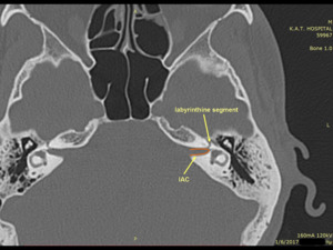
Fig. 5: The labyrinthine segment.
References: Department of Radiology, KAT General Hospital, Athens, Greece
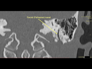
Fig. 6: The facial (Fallopian) canal (bony canal from the IAC to the stylomastoid foramen).
References: Department of Radiology, KAT General Hospital, Athens, Greece
The tympanic segment (geniculate ganglion to pyramidal eminence).
(Figure 7)
At the geniculate ganglion makes the facial nerve a 75 degree turn posteriorly to become the tympanic segment.
Within the tympanic cavity,
the facial nerve passes medial to the incus.
It runs posterior-superior to the cochleariform process,
superior and lateral to the oval window,
and then inferior to the lateral semicircular canal.
At the pyramidal process,
the tympanic segment turns inferiorly at a 95-125 angle (at the second genu) to become the mastoid segment.
The tympanic segment has no branches.
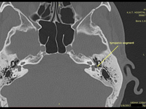
Fig. 7: The tympanic segment.
References: Department of Radiology, KAT General Hospital, Athens, Greece
The mastoid segment (pyramidal eminence to stylomastoid foramen).
(Figure 8)
The mastoid segment of the facial nerve runs posteromedially along the external auditory canal to its exit from the temporal bone at the stylomastoid foramen.
Two branches arise from the mastoid segment:
- the nerve to the stapedius
- the chorda tympani
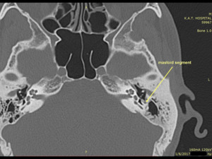
Fig. 8: The mastoid segment.
References: Department of Radiology, KAT General Hospital, Athens, Greece
The extratemporal/extracranial segment (from stylomastoid foramen to post parotid branches).
The nerve exits the temporal bone at the stylomastoid foramen.
It gives off the posterior auricular nerve and then gives off two small motor nerves to the stylohyoid muscle and the posterior belly of the digastrics muscle,
which are typically below the resolution of MRI.
After the facial nerve crosses lateral to the styloid process,
it dives into the parotid gland,
where it follows a virtual plane between the deep and superficial lobes of the parotid gland and splits into a nerve plexus called the pes anserinus (“goosefoot”),
which represents a convenient surgical landmark.
At the pes anserinus,
the facial nerve subdivides into 5 terminal branches:
- Temporal nerve
- Zygomatic nerve
- Buccal nerve
- Marginal mandibular nerve
- Cervical nerve
Important:
- The facial nerve is the only cranial nerve that may show normal post-contrast enhancement (in the majority of the cases is asymmetric left-to-right).
- No enhancement should be seen in:
- Cisternal segment
- Meatal segment
- Extratemporal/extracranial segment
- CT is preferable for imaging the lateral course of the facial nerve from the porus acusticus to the stylomastoid foramen.
- Facial nerve cannot be detected within the parotid gland with conventional imaging.

 Anatomy of the Human Body. Gray's Anatomy, Plate 788")
 Illustration of the facial nerve nuclei. (B) MRI axial T2 showing the loop of the motor fibres of the facial nerve posteriorly over the abducens nerve. References: Richard S. Snell Clinical Neuroanatomy (7th Ed.), Radiopaedia (Case courtesy of A.Prof Frank Gaillard, Radiopaedia.org, rID: 50811),")

 The diagram for the orientation of the nerves of the internal acoustic meatus. (B) The annotated sagitally T2-weighted, thin-slice MR image of the internal auditory canal showing the facial nerve situated at the anterior superior quadrant of the internal auditory canal. (VII = facial nerve, NI = nervus intermedius, VIIIc = cochlear nerve, VIIIvs = superior division of vestibular nerve, VIIIvi = inferior division of vestibular nerve) References: Radiopaedia (Case courtesy of A.Prof Frank Gaillard, Radiopaedia.org, rID: 36049), Department of Radiology, KAT General Hospital, Athens, Greece")

 canal (bony canal from the IAC to the stylomastoid foramen). References: Department of Radiology, KAT General Hospital, Athens, Greece")

