Type:
Educational Exhibit
Keywords:
Abdomen, Gastrointestinal tract, Anatomy, CT, MR, Fluoroscopy, Contrast agent-oral, Diverticula
Authors:
N. Kinger1, P. Mittal2; 1Atlanta, GA/US, 2Decatur, GA/US
DOI:
10.26044/ecr2019/C-0707
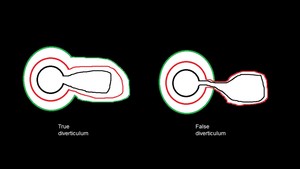
Background
A diverticulum is defined an outpouching of a hollow organ.
The entirety of the gastrointestinal tract can be affected by varying numbers and types of diverticula.
These diverticula,
including upper tract and lower tract diverticula,
can be sources of significant morbidity and even mortality through complications such as bleeding,
infection,
fistula formation,
and perforation.
Gastrointestinal diverticula can be subdivided into true and false diverticula and also acquired and congenital diverticula.
A true diverticulum contains all layers of the organ wall.
A false diverticulum does not contain all layers of the organ wall and generally includes mucosa and submucosa with herniation through the muscular layer.
Fig. 1: True versus False Diverticulum
Upper gastrointestinal diverticula,
those ranging from mouth through proximal portions of the small bowel,
can be well evaluated with fluoroscopic imaging with CT allowing for identification of complications particularly in the small bowel.
Lower gastrointestinal diverticula from distal small bowel through the rectum are often well evaluated with CT followed by MRI and fluoroscopy as indicated.