ECR 2019 / C-1795
Minimally invasive extraction of foreign bodies by USG and fluoroscopy
Congress:
ECR 2019
Poster Number:
C-1795
Type:
Educational Exhibit
Keywords:
Foreign bodies, Education and training, Artifacts, Treatment effects, Surgery, Removal, Ultrasound, Fluoroscopy, Musculoskeletal soft tissue, Interventional non-vascular, Extremities
Authors:
J. Murillo1, C. RIVERA2; 1Tegucigalpa, FM/HN, 2Tegucigalpa,D.C./HN
DOI:
10.26044/ecr2019/C-1795

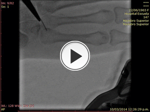
Fig. 1

Fig. 2:
RX. Simple with projections A. Oblique and B. Lateral. Showing radio opaque...

Fig. 3:
US showing hyperechoic linear image of 27 mm in length, in soft tissue in the...

Fig. 4:
Multiple images, extraction a foreign body guided by US. Minimal soft tissue...

Fig. 5:
Multiple images of Rx. Simple and US, sequential photography of event...

Fig. 6:
RX. Simple with multiple projections of the right hand, with a radio opaque...

Fig. 7:
Sequential images of Fluroscopy when extracting the foreign body.

Fig. 9:
Minimal soft tissue lesion when extraction guided by FLUROSCOPY

Fig. 10:
Multiple images of Rx. simple, fluoroscopy and sequential photography of event...