Keywords:
Pathology, Neoplasia, Education and training, Surgery, Diagnostic procedure, MR, Neuroradiology brain, Head and neck, CNS
Authors:
T. M. B. Da Concei├¦├Żo1, J. Quedi Ara├║jo da Silva2, ’┐Į. C. Martins Antunes 2, M. Muxfeldt Bianchin 2, J. ├üvila Duarte2; 1Porto Alegre, RS/BR, 2Porto Alegre/BR
DOI:
10.26044/ecr2019/C-1871
Methods and materials
Patients and Methods
A total of forty-five patients who were diagnosed as macroadenomas (size > 1.0 cm) and admitted to our hospital between January 2010 and January 2018 were included in this study.
All patients underwent a transsphenoidal operation by a single surgeon,
who determined during the surgery the consistency of PAMs,
classifying them in accordance with its consistency in soft or fibrous.
Adenomas were classified of soft consistency if the tumor could be easily removed with curettage and aspiration.
On the contrary,
adenomas were classified in fibrous if the tumor could not be suctioned with an aspirator,
and bipolar electrocoagulation or sharp segmentation resection was needed.
This definition soft or fibrous is rather subjective but it has been widely used by the operating surgeon during surgical procedures.
Neuroimaging
MR imaging was performed using a 1.5 Tesla Achieva MR scanner (41 patients) or 3.0 Tesla Ingenia MR scanner (4 patients),
both scanners were from PhilipsŌōć Medical Systems,
Best,
Netherlands.
Utilizing the MR T2WI sequence,
a ratio was calculated using PAMs signal and the cerebellar peduncle measurements,
as proposed by Smith et al [1].
All neuroimaging and T2WI sequence ROIs (region of interest) of adenoma,
cerebellar peduncle and adenoma- to-cerebellar peduncle ratios(ACP) were calculated independently by three board certificated radiologists,
that were blinded to the perception of intraoperative consistency by the neurosurgeon and to the histopathological diagnosis.
When results differ among radiologists,
a consensus was obtained.
An ROI was manually traced on T2WI using an electronic cursor.
ROI measurements were obtained from the solid component of the tumoral lesions with the lowest signal intensity on T2WI,
excluding necrotic and hemorrhagic areas.
The ROIŌĆÖs were selected within the adenoma for a representative sampling in homogenous intensity and an average intensity value was calculated for each tumor.
At the second step,
the ROI was chosen from the cerebellar peduncle as a standard anatomical denominator that can be easily selected due to homogeneously MR SI and the reduced error fig-1a,
1b).
The other imaging characteristics,
such as the SI,
the tumor contrast enhancement,
shape or morphology of lesions were not considered in this study.
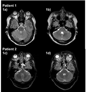
Fig. 1: Fig 1a-1b). Patient number 01, the adenoma-cerebellar ratio (ACP). 1a) ROI in the homogeneous macroadenoma MRI T2-weighted sequence. 1b) ROI in the middle peduncle on MRI T2-weighted sequence. Fig 1c-1d. Patient number 02, the adenoma-cerebellar ratio (ACP). 1c) An example of ROI in the heterogeneous macroadenoma MRI T2-weighted sequence and 1d) ROI in the middle peduncle on MRI T2-weighted.
