ECR 2019 / C-2497
Can apparent diffusion coefficient (ADC) values improve the diagnosis of peripheral zone prostate cancer (PZPCa) in PIRADSv2 score 4?
Congress:
ECR 2019
Poster Number:
C-2497
Type:
Scientific Exhibit
Keywords:
Genital / Reproductive system male, Oncology, MR-Diffusion/Perfusion, Diagnostic procedure, Biopsy, Cancer, Outcomes
Authors:
S. Verna1, S. Agostini1, E. Bertelli1, F. Parretti1, S. Lucarini1, F. De Nisco1, M. Mastrorosato1, L. N. Mazzoni2, V. Miele1; 1Florence/IT, 2Pistoia/IT
DOI:
10.26044/ecr2019/C-2497
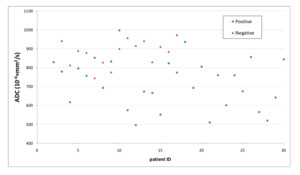
 scatterplot of peripheral zone lesions classified as PIRADSv2 4, respectively positive and negative for prostate cancer at biopsy. It can be seen that tissues positive for prostate cancer show lower ADC values.")
Fig. 4:
Apparent Diffusion Coefficient (ADC) scatterplot of peripheral zone lesions...
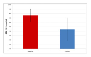
 histogram (mean±standard deviation) of peripheral zone lesions with PIRADSv2 4, respectively positive and negative for prostate cancer at biopsy. It can be seen that tissues positive for prostate cancer show lower ADC values.")
Fig. 5:
Apparent Diffusion Coefficient (ADC) histogram (mean±standard deviation) of...