Type:
Educational Exhibit
Keywords:
Musculoskeletal joint, Musculoskeletal bone, Trauma, CT, Conventional radiography, MR, Education
Authors:
F. G. Rodriguez-Ruiz1, A. Saldana2, E. Trullenque3, L. M. Montes Chinea4; 1Caguas, Puerto Rico/US, 2San Juan, Puerto Rico/US, 3San Juan, PR/US, 4San Juan/US
DOI:
10.26044/ecr2019/C-3513
Background
I.
Introduction
The wrist is a complex joint that forms the bridge between the forearm and hand providing stability,
control and coordinating the movements.
The wrist is composed of eight carpal bones that articulate with the distal radio-ulnar joint (DRUJ) proximally and the base of the metacarpals distally.
The wrist igaments are categorized as intrinsic or as extrinsic.
II.
Anatomy
A.
Carpal Bones
- There are two rows of carpal bones.
The proximal row articulate with the DRUJ,
contains the scaphoid,
lunate,
triquetrum and pisiform.
It plays an important role in coordinating the movements and controlling the forces transmitted from the forearm to the arm 11.
- The distal row contains the trapezium,
trapezoid,
capitate and hamate.
It is responsible for providing the wrist stability and articulating with the metacarpals.
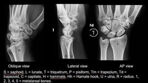
Fig. 1
References: Department of Diagnostic Radiology, University of Puerto Rico
B.
Ligaments
The intrinsic ligaments connect adjacent carpal bones.
Extrinsic ligaments are responsible for joining the radius and ulna to the carpal bones.
Radial and ulnar collateral ligaments
The radial collateral ligament is located between the styloid process of the radius and the scaphoid bone.
The ulnar collateral ligament,
between the styloid process of the ulna and the triquetrum and pisiform bones.
Dorsal ligaments
Are weaker and less important than the volar ligaments.
The two most important dorsal ligaments are the dorsal intercarpal ligament and the dorsal radiotriquetral ligament.
Palmar or Volar Ligaments
Join the different carpal bones to the radius and the ulna.
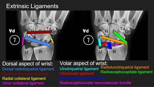
Fig. 2
References: Department of Diagnostic Radiology, University of Puerto Rico
III.
Imaging Modalities for Evaluation of the Wrist
When evaluating the wrist in a patient that fell on an outstretched hand (FOOH),
the wrist plain films are the initial imaging modality used to assess the wrist.
The evaluation should include at least 2 views [Anterior-Posterior (AP) and lateral views].
Some carpal fractures are difficult to see on conventional radiograph due to the superimposition of the carpal bones and soft tissue on lateral views.
The referring clinician’s level of suspicion in a wrist fracture is very important to not delay the diagnosis and management of carpal fractures.
Computer Tomography (CT) and Magnetic Resonance Imaging (MRI) are increasing their role on evaluating for acute wrist trauma when: there is clinical suspicion of a carpal fracture with negative plain films,
initial radiographs are indeterminate or for surgical planning11.
