ECR 2020 / C-04208
Computed Tomography Findings with Histopathological Correlation in Inflammatory, Benign & Malignant Appendiceal Mucoceles
Congress:
ECR 2020
Poster Number:
C-04208
Type:
Scientific Exhibit
Keywords:
Abdominal Viscera, Abdomen, Gastrointestinal tract, CT, Biopsy, Surgery, Inflammation, Neoplasia, Pathology, Retrospective, Cross-sectional study, Performed at one institution
Authors:
K. Khandwala1, N. Sajjad2, W. A. Memon1, A. Malik3; 1Karachi/PK, 2Karachi, Si/PK, 3Atlanta/US
DOI:
10.26044/ecr2020/C-04208
.")
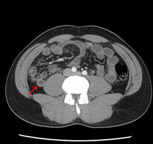
Fig. 1:
Appendiceal mucocele with co-existing appendicitis in a 35-year-old male with...
. On pathology, this was a low-grade mucinous neoplasm.")
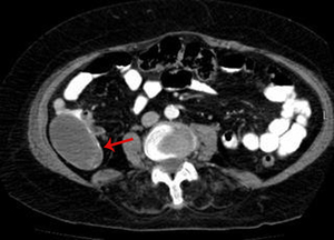
Fig. 2:
Appendiceal mucocele in an 83 year old female with vague abdominal pain. Mural...

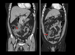
Fig. 3:
Appendiceal mucocele in a 57 year old male with abdominal distension. Soft...

Fig. 4:
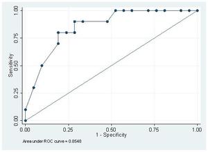
ROC curve analysis for inflammatory versus neoplastic mucoceles against...