ECR 2020 / C-10677
Key points in computed tomographic (CT) evaluation of perirenal transplant fluid collections
Congress:
ECR 2020
Poster Number:
C-10677
Type:
Educational Exhibit
Keywords:
Observational, Retrospective, Transplantation, Haemorrhage, Abscess, Complications, CT, Urinary Tract / Bladder, Kidney, Genitourinary, Performed at one institution
Authors:
I. A. Cristache1, M. Buzoianu2, R. A. Capsa1, M. Grasu2, I. G. Lupescu2; 1Bucuresti/RO, 2Bucharest/RO
DOI:
10.26044/ecr2020/C-10677
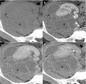
 and enhanced CT in cortico-medullary phase (b), nephrogenic phase (c) and excretory phase (d): large perirenal hyperdense nonenhancing collection (*) compressing the renal parenchyma and sinus. References: Radiology, Medical Imaging and Interventional Radiology of Fundeni Clinical Institute, Bucharest, Romania")
Fig. 1:
Perirenal graft hematoma. Unenhanced (a) and enhanced CT in cortico-medullary...
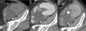
 and enhanced CT in nephrogenic phase (b) and excretory phase (c). Perirenal graft lympocele: small perirenal fluid nonenhancing collection (arrow).
References: Radiology, Medical Imaging and Interventional Radiology of Fundeni Clinical Institute, Bucharest, Romania")
Fig. 2:
Perirenal graft lymphocele. Unenhanced (a) and enhanced CT in nephrogenic phase...
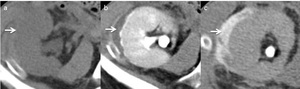
 and enhanced CT evaluation in early excretory phase (b) and late excretory phase (c) of a small perirenal collection (arrow) progressively filling with contrast material.
References: Radiology, Medical Imaging and Interventional Radiology of Fundeni Clinical Institute, Bucharest, Romania")
Fig. 3:
Perirenal graft urinoma. Unenhanced (a) and enhanced CT evaluation in early...

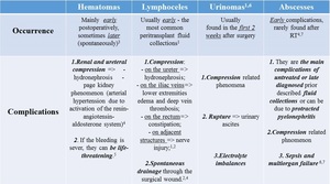
Table 1:
Features of perigraft fluid collections