ECR 2020 / C-10859
Is there bowel involvement? : A comparison of local protocols against ESUR Guidelines for imaging of endometriosis
Congress:
ECR 2020
Poster Number:
C-10859
Type:
Scientific Exhibit
Keywords:
Genitourinary, Colon, Contrast agents, Genital / Reproductive system female, MR, Contrast agent-other, Health policy and practice, Outcomes analysis, Outcomes, Retrospective, Diagnostic or prognostic study, Performed at one institution
Authors:
R.-E. Chung1, S. LEE2, A. Stankiewicz1, J. ritchie1; 1Stoke-on-Trent/UK, 2Stoke on Trent/UK
DOI:
10.26044/ecr2020/C-10859
 MRI findings in endometriosis :https://posterng.netkey.at/esr/viewing/index.php?module=viewing_poster&task=viewsection&pi=128262&ti=428336&si=1482&searchkey=")
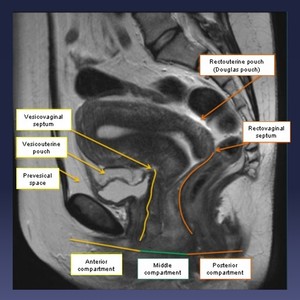
Fig. 1:
Figure 1. MRI Pelvis T1w Sagittal view highlighting the normal anatomy.
 guidelines: MR imaging of pelvic endometriosis Eur Radiol (2017) 27:2765–2775 DOI 10.1007/s00330-016-4673-z 5 December 2016")
Fig. 2:
Guidelines published by ESUR on MRI imaging of pelvic endometriosis.