ECR 2020 / C-10892
Role of Dual Energy Computed Tomography (DECT) in evaluation of Renal stones: What a surgeon wants to know?
Congress:
ECR 2020
Poster Number:
C-10892
Type:
Educational Exhibit
Keywords:
Not applicable, Calcifications / Calculi, Surgery, Lithotripsy, CT, Urinary Tract / Bladder, Kidney, Genitourinary
Authors:
A. Kanwar, S. Mehra; New Delhi/IN
DOI:
10.26044/ecr2020/C-10892

Fig. 4:
Scout image for Non contrast CT to be acquired initially to localize renal...

Fig. 5:
NCCT showing location of renal stone

Fig. 6:
Targeted Dual energy scanning done at region of renal stone
")
Fig. 7:
Targeted Dual energy scanning done at region of renal stone(sagittal section)

Fig. 11:
NCCT for a renal stone of DE ratio 1.41, calcium based stone

Fig. 12:
DECT for a renal stone of DE ratio 1.41, calcium based stone

Fig. 13:
DE ratio graph for a renal stone of DE ratio 1.41, calcium based stone

Fig. 8:
NCCT for a renal stone with DE ratio 1.44 i.e. calcium based stone

Fig. 9:
DECT for a renal stone with DE ratio 1.44 i.e. calcium based stone

Fig. 10:
DE ratio graph for a renal stone with DE ratio 1.44 i.e. calcium based stone

Fig. 14:
NCCT of a renal calculi with DE ratio 1.22, Cystine based stone

Fig. 15:
DECT of a renal calculi with DE ratio 1.22, Cystine based stone

Fig. 16:
DE Ratio graph of a renal calculi with DE ratio 1.22, Cystine based stone

Fig. 17:
Measuring renal stone to assess stone burden

Fig. 18:
Number and location of stones should be mentioned in case of multiple calculi

Fig. 19:
Lower calyx stone at L2 level
")
Fig. 20:
Dilated pelvicalyceal system with adjacent perinephric fat stranding (signs of...

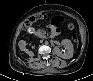
Fig. 21:
Stone to skin distance measured laterally.

Fig. 22:
Stone to skin distance measured posteriorly.

Fig. 23:
Cortical thickness at the level of stone

Fig. 24:
Pre surgical planning on coronal reformatted CT image to ascertain safe path...

Fig. 25:
Thinned out renal cortex with adjacent perinephric fat stranding.
PCNL tube...