Spinal Lipoma Classification
The diagnosis and classification of spinal lipomas were made on the basis of T1- and T2-weighted MRI using mid- and parasagittal views.
Type 1: Primary Neurulation Failure Only
Type 1 spinal lipoma is a typical form in which the lipoma-cord interface occurs on the dorsal surface of the spinal cord, while the conus medullaris is located distally or ventrally with respect to the caudal end of the lipoma. The subcutaneous fat mass penetrates the fascia and reaches the dorsal surface of the dura through the pathological spina bifida.The intradural lipoma is usually located in the dorsal or dorsolateral aspect of the spinal cord, which is tethered caudally (Fig.6).
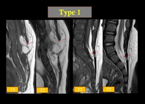
Fig. 6: Type 1- primary neurulation failure.
Lipoma cord interface seen dorsal surface of the spinal cord.
Conus medullaris seen ventral to the caudal end of intraspinal lipoma.
Type 2: Failed Neurulation Between the Primary and Secondary Stages
Type 2 spinal lipoma also penetrates the fascia and continues into the intraspinal lesion through the pathological spina bifida. The lipoma is attached to the spinal cord, but the conus medullaris is malformed and difficult to recognize by MRI . The difference between Types 1 and 2 is determined by the presence or absence of the conus medullaris. Because Type 1 spinal lipomas are formed purely by failed primary neurulation, the conus medullaris is present caudally or ventrally with respect to the spinal lipoma. The tapered shape of the end of the spinal cord suggests the presence of the conus medullaris . On the other hand, the Type 2 spinal lipoma is the result of failed neurulation between the primary and secondary stages. The end of the spinal cord where the conus medullaris should exist is undifferentiated and merges with the spinal lipoma. A sharp tapered end to the spinal cord is difficult to confirm in Type 2 spinal lipomas(Fig .7&8).
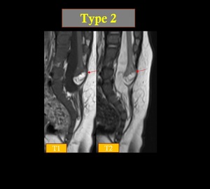
Fig. 7: Conus ends at L5 vertebral level tethered by terminal lipoma which is continuous with subcutaneous fat through the defect in the posterior elements of L5 vertebrae, more on the left side.
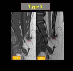
Fig. 8: spinal lipoma seen caudal end of spinal canal with posterior dural defect & spina bifida & extension into subcutaneous plane. Cord seen upto S2 vertebrae level. Caudal end of spinal cord fused to the spinal lipoma without obvious conus medullaris formation & without clear border line.
Type 3: Early Phase Secondary Neurulation Failure
This type of spinal lipoma involves the conus medullaris, the caudal end of which is unformed and directly connects to the spinal lipoma. The lipoma extends in the caudal direction, penetrating the caudal end of the dura to connect with subcutaneous fat through the enlarged sacral hiatus. The dorsal surface of the dura remains intact. In contrast to Type 2, in Type 3 spinal lipomas there is no pathological spina bifida or fascial defect due to the lipoma (Fig.9).
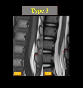
Fig. 9: Conus medularis incompletely formed and fuses with caudally located lipoma but no clear borderline.There is no dural defect on dorsal side.
Type 4: Late Phase Secondary Neurulation Failure
In this type, the spinal lipoma is located in the filum terminale. The caudal end of the conus medullaris is easily confirmed on MRI, and most or part of the filum terminale is composed of a linear string-like lipoma(Fig 10&11).
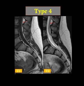
Fig. 10: Filum terminal lipoma seen at L3 to S2 level
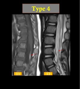
Fig. 11: Filum terminal lipoma noted at L2-L3 level,normal conus medularis
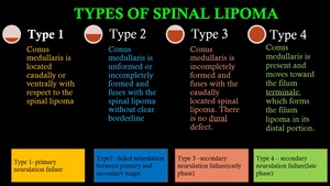
Fig. 12
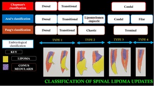
Fig. 13
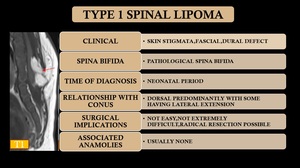
Fig. 14
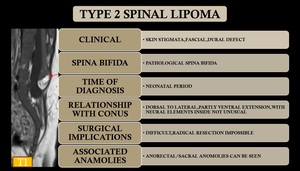
Fig. 15
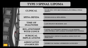
Fig. 16
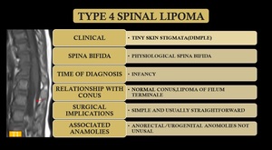
Fig. 17
