Keywords:
Abdomen, Liver, Ultrasound physics, Ultrasound, Diagnostic procedure, Equipment, Cirrhosis, Image registration, Tissue characterisation
Authors:
Y. Solskaya, M. Tirane, P. Prieditis, A. Staka, A. Upena, L. Saule, M. Radzina; Riga/LV
DOI:
10.26044/ecr2021/C-12553
Methods and materials
Total 154 patients were enrolled in a prospective study between April 2019 and December 2020, where 98 patients with the confirmed diffuse liver disease, 17 healthy patients (63 women and 52 men, median age 52.0) and additionally 56 patients with proven SARS-CoV-19.
Patients with the severe liver parenchymal changes due to rare disease as Budd-Chiari and Wilson disease were excluded.
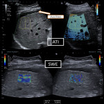
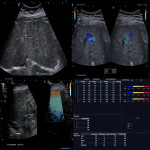
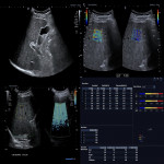
For fibrosis, steatosis detection and viscosity quantification the multiparametric measurements were performed by three radiologists, experienced in abdominal ultrasound: B-mode imaging, 2D shear-wave elastography measurements, attenuation imaging (ATI) and dispersion imaging on Ultrasound machine Aplio i800, Canon Medical Systems, Japan.
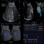
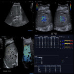
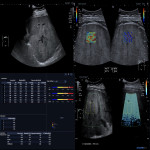
Average 5-13 (+/-2.3 SD) numbers of Elasto - measurements in 2D-SWE were registered with IQR/median values (Figure 1),
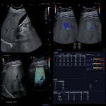
including patients with ascites (Figure 2).
The laboratory results as ALAT and ASAT were taken for a correlation.
Chronic liver parenchymal changes were caused by different etiology - virus hepatitis B (n=3), hepatitis C (n=31), autoimmune hepatitis (n=6), steatohepatosis (n=37), alcohol related (n=13) and patients of other etiology (n=8).
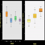
Patients with confirmed SARS-CoV-19 infection in the last 3-9 months were scanned. Multiparametric ultrasound measurements were performed with the liver biochemical damage and inflammation marker control (LDH, GGT, AlAT, CRP, ESR), where in higher grade of steatosis a more severe disease course was found.

