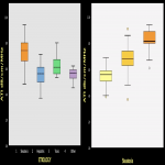
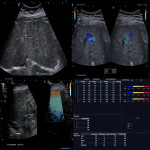
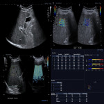
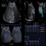
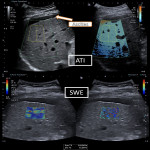
There was a statistically significant difference among etiology groups and ATI (p=0.0001) with predominantly high attenuation intensity range dB/cm/MHz >0.65-0.8 in steatohepatosis (strong correlation r=0.71; p=0.0001).
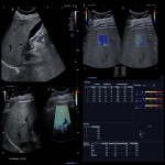
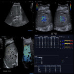
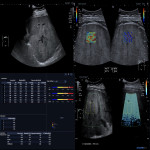
Relatively high attenuation intensity range was detected in toxic hepatitis group up to 0.55-0.65 dB/cm/MHz with steatosis qualitative assessment on B mode image (normal or hyperechogenic liver structure) (rs=0.5; p=0.0001) (Figure 3, Figure 4, Figure 5).
Alterations in liver tissue viscosity to the stage of fibrosis were revealed with positive correlation (rp=0.40; p=0.005) with higher value towards higher fibrosis stage, but no correlation to steatosis was found (p=0.051).
Attenuation (ATI) median values had moderate correlation with steatosis qualitative assessment on B mode image (normal or hyperechogenic liver structure) (rs=0.5; p=0.0001) (Figure 6, Figure 7, Figure 8).
Post Covid-19 patients showed the higher dispersion values (median 12.4 (m/s)/KHz) as moderate viscosity (50%) with reference of <12 (m/s)/KHz as normal values as well as no changes in ATI results [6]. It is possible that shear wave dispersion alone may provide interesting information on tissue organization at the microscopic level and give information on necro-inflammatory activity.
A third part of study group of Covid-19 patients have been hospitalised and in cases of severe disease - the steatosis grades were significantly higher (F= 9.1, p<0.01) after 3-6 months after onset.
In Covid-19 patients with no previous history of liver disease 2D shear wave results showed F0-F1 stage of fibrosis, that indicates initial changes.
According to the results of our study and analyzing the study performed in 2017 by F.Piscaglia et al. [7] multiparametric ultrasound approach can be a good non-invasive measurement tool for evaluating the stage of liver fibrosis and inflammatory activity. The accuracy of the method is examinator dependent, although some limitations as ascites in big volume or patient non-contribution may be present.
In our study we didn’t find the correlation of the stage of fibrosis to the degree of steatosis, also this effect was not confirmed during the previous studies as Bota et al. 2011 [8] and J.Yoo et al.2019 [9].
The results of our study showed that laboratory confirmed inflammatory changes in liver gave higher numbers in dispersion and therefore in viscoelasticity.
Further research is required with the liver tissue biopsy, to detect the relationship between the fibrosis, viscosity and pathological changes.

