In general,
a complaint of back pain in a child is considered uncommon.
But the reality is different.
By the age of 15,
20-70% of children will report back pain.
When a body is acted upon by gravity,
all the mass particles of which the body is composed experience a force of attraction directed toward the Earth's center.
The resultant force of all of these small attractive forces is the body's weight and the location at which the resultant force is assumed to act is the center of gravity of the body [1].
Adult man,
of all animals,
including mammals,
is the only exclusively bipedal.
The upright man is extremely unstable,
its center of gravity is too high.
We are only stable if the center of gravity is located over the base of support of both feet.
This requires a complex and permanent neuromuscular control.
Fig. 2
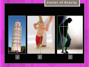
Fig. 2: Perfect Balance: The vertical line through the center of gravity should cut the base lift both feet (Fig.A,C). In image B the feet and the center of gravity are not aligned and a support is needed to maintain the balance
References: Hospital of Galdácano. Radiology
A child will take approximately twelve months to acquire the necesary control to move without falling and also to acquire the normal sagittal profile [2].
At birth a C-shaped vertebral column is seen.
As a neonate starts lifting its head,
a cervical curvature begins to develop.
As the infant begins to crawl,
then walk and assumes an upright posture,
the lumbar curvature forms.
Fig. 3
The thoracic and sacral kyphosis are primary curves because they were present at birth.
The cervical and lumbar lordosis are secondary curves.
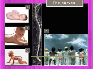
Fig. 3: Development of spinal curves. C-shaped vertebral (A). The cervical curvature begins to develop (B.The lumbar curvature forms (C). Center of gravity (D).
References: Hospital of Galdacano. Radiology
Sagittal Balance: Sagittal balance refers to the position of the head in relation to the pelvis.
Normal sagittal balance is essential for the ability of the individual to stand in the upright position with minimal effort.
Radiographic assessment is done using a standing full-length lateral radiograph obtained with extension at the hips and knees.
The C7 vertebral body and the hip joint should be visualized.
A line is drawn perpendicular to the ground (plumb line) from the center of the C7 vertebral body to the pelvis.
The distance of this line to the posterosuperior aspect of the S1 vertebral body is the sagittal vertical axis offset.
The normal range for the sagittal vertical axis offset may be anywhere between 2.5 cm and 5.0 cm.
Fig. 4
- Negative sagittal imbalance: Plumb line falls behind the posterosuperior corner of the S1 vertebral body.
- Neutral sagittal balance: Plumb line intersects the posterosuperior corner of the S1 vertebral body.
- Positive sagittal imbalance: Plumb line falls in front of the posterosuperior corner of the S1 vertebral body.
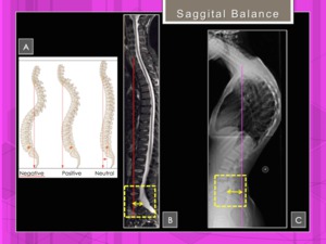
Fig. 4: Sagittal balance or plump line. Neutral sagittal balance: Plumb line intersects the posterosuperior corner of the S1 vertebral body (B). Negative sagittal imbalance: Plumb line falls behind the posterosuperior corner of the S1 vertebral body (C).
References: Hospital of Galdácano. Radiology
Each segment of the spine is designed for a certain range of motion.
Observed variations in the curves and spinal angle values confirm the physiological changes produced by the transition from supine to orthostatic position.
Fig. 5
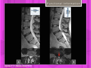
Fig. 5: Functional information. This spinal instability was visible only when the patient was scanned upright(B), and undetectable on a conventional, lie-down MRI scanner (A).
References: Hospital of Galdácano. Radiology
Pain symptoms in the adolescent can often arise from the postural changes.
Approximately 25-40% of children will have alterations in imaging studies that indicate a pathological cause for their back pain.
In this study we exclude congenital disorders,
neuromuscular problems,
traumatic injuries,
tumors,
infections and arthritic disorders.
Our main topics are:
- Scheuermann's Kyphosis
- Scoliosis
- Spondylolisthesis (+/- Spondylolisis)
The radiologist will diagnose these conditions using also new MRI imaging options.

. In image B the feet and the center of gravity are not aligned and a support is needed to maintain the balance References: Hospital of Galdácano. Radiology")
. The cervical curvature begins to develop (B.The lumbar curvature forms (C). Center of gravity (D). References: Hospital of Galdacano. Radiology")
. Negative sagittal imbalance: Plumb line falls behind the posterosuperior corner of the S1 vertebral body (C). References: Hospital of Galdácano. Radiology")
, and undetectable on a conventional, lie-down MRI scanner (A). References: Hospital of Galdácano. Radiology")