ESSR 2016 / P-0120
It’s normal, but it hurts! Painful sesamoid and accessory bone syndromes of the foot.
Congress:
ESSR 2016
Poster Number:
P-0120
Type:
Educational Poster
Keywords:
Pathology, Normal variants, Education, Diagnostic procedure, MR, CT, Conventional radiography, Musculoskeletal bone, Bones, Anatomy
Authors:
A. C. Vieira, A. Vieira, R. Cunha; Porto/PT
DOI:
10.1594/essr2016/P-0120
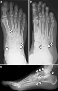
, oblique (b) and lateral (c) - 1 Os trigonum, 2 os peroneum, 3 os naviculare, 4 os intermetatarseum, 5 os vesalianum 6 os supranaviculare, 7 os supratalare, 8 os talotibiale, 9 os calcaneus secundarium References: Insights Imaging (2013) 4:581–593")
Fig. 1:
Acessory bones of the foot - AP (a), oblique (b) and lateral (c) - 1 Os...

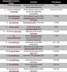
Table 1:
Accessory and sesamoid bones: locations and prevalence