ESSR 2017 / P-0282
3D printing (3DP) applications in complex revision hip arthroplasty (CRHA): A prospective nonrandomised controlled observational cohort study.
Congress:
ESSR 2017
Poster Number:
P-0282
Type:
Scientific Poster
Keywords:
Anatomy, Computer applications, Bones, CT, Image manipulation / Reconstruction, CT-Quantitative, Computer Applications-3D, CAD, Technical aspects, Arthritides, Outcomes
Authors:
D. Dalili, N. Byrne, Z. Shah, M. J. Bankes, M. George, A. Isaac; London/UK
DOI:
10.1594/essr2017/P-0282

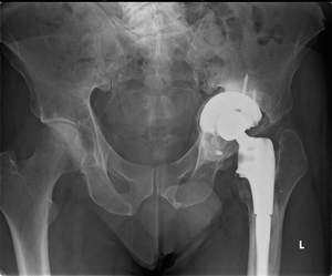
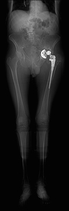
Fig. 1:
Patient 1 - preoperative AP pelvic radiograph

Fig. 2:
Patient 1 - preoperative AP left hip radiograph

Fig. 3:
Patient 1 - preoperative lateral hip radiograph

Fig. 4:
Patient 1 preoperative plain radiograph of the left hip joint.
The patient...

Fig. 5:
Patient 1 preoperative plain radiograph of the left hip joint.
The patient...

Fig. 6:
Patient 1 preoperative lateral plain radiograph of the left hip joint.
The...

Fig. 10:
Patient 1 - CT scout, demonstrating limb length discrepancy, pivotal to...
 References: Amanda Isaac")
Fig. 12:
Patient 1 - 3D printed model 1:1 scale (true to size)
 References: Amanda Isaac")
Fig. 13:
Patient 1 - 3D printed model 1:1 scale (true to size)
 References: Amanda Isaac")
Fig. 14:
Patient 1 - 3D printed model 1:1 scale (true to size)
 References: Amanda Isaac")
Fig. 15:
Patient 1 - 3D printed model 1:1 scale (true to size)
 References: Amanda Isaac")
Fig. 16:
Patient 1 - 3D printed model 1:1 scale (true to size)

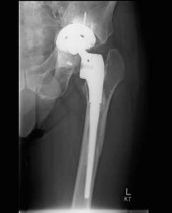
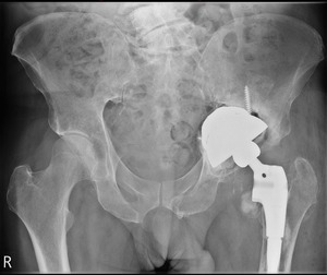
Fig. 17:
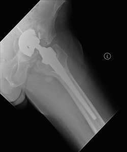
Patient 1 - post revision AP pelvic radiograph

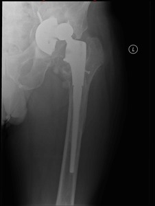
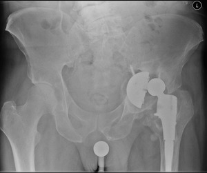
Fig. 18:
Patient 2 - preoperative AP pelvic radiograph

Fig. 19:
Patient 2 - preoperative AP hip radiograph

Fig. 20:
Patient 2 - preoperative lateral hip radiograph

Fig. 21:
Patient 2 - intra-operative hip aspiration to exclude infection

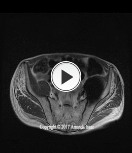
Fig. 25:
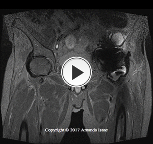
Patient 2 - volume rendered reformats. An attempt to visualise the pelvis and...

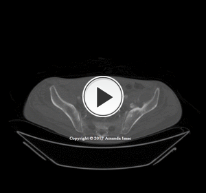
Fig. 26:
Patient 2 - volume rendered reformats. An attempt to visualise the pelvis and...

Fig. 27:
Patient 2 - volume rendering reformats. An attempt to visualise the pelvis and...

Fig. 28:
Patient 2 - volume rendering reformats. An attempt to visualise the pelvis and...

Fig. 29:
Patient 2 - volume rendering reformats. An attempt to visualise the pelvis and...

Fig. 30:
Patient 2 - volume rendering reformats. An attempt to visualise the pelvis and...

Fig. 31:
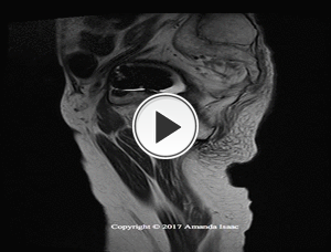
Patient 2 - Software analysis for 3D printing

Fig. 32:
Patient 2 - Software analysis for 3D printing - small FOV

Fig. 33:
Patient 2 - Software analysis for 3D printing. Subtracted femoral components...

Fig. 34:
Patient 2 - 3D Planning oblique. Granulation tissue subtracted to assess bone...

Fig. 35:
Patient 2 - 3D Planning large FOV. Granulation tissue subtracted to assess bone...

Fig. 36:
Patient 2 - 3D Planning PA. Granulation tissue subtracted to assess bone...

Fig. 37:
Patient 2 - 3D Planning small FOV. Granulation tissue subtracted to assess bone...
 References: Amanda Isaac")
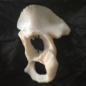
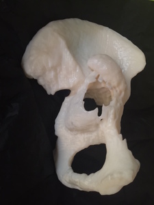
Fig. 38:
Patient 3 - 3D printed model (1:1 scale)
 References: Amanda Isaac")
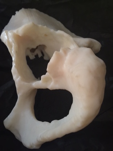
Fig. 39:
Patient 3 - 3D printed model (1:1 scale)
 - Acetabular cup References: Amanda Isaac")
Fig. 40:
Patient 3 - 3D printed model (1:1 scale) - Acetabular cup
. Acetabular depth References: Amanda Isaac")
Fig. 41:
Patient 3 - 3D printed model (1:1 scale). Acetabular depth
 References: Amanda Isaac")
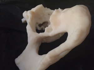
Fig. 42:
Patient 4 - 3D printed model (1:1 scale)
. Acetabular cup References: Amanda Isaac")
Fig. 43:
Patient 4 - 3D printed model (1:1 scale). Acetabular cup
. Lateral oblique References: Amanda Isaac")
Fig. 44:
Patient 4 - 3D printed model (1:1 scale). Lateral oblique

Fig. 50:
Thickness map for 3D printed model

Fig. 51:
Thickness map for 3D printed model

Fig. 52:
Thin bone mesh

Fig. 53:
Thin bone mesh
 References: Amanda Isaac")
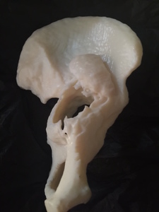
Fig. 45:
Patient 5 - 3D printed model (1:1 scale)
. Acetabulum References: Amanda Isaac")
Fig. 46:
Patient 5 - 3D printed model (1:1 scale). Acetabulum
. Bone loss oblique References: Amanda Isaac")
Fig. 47:
Patient 5 - 3D printed model (1:1 scale). Bone loss oblique

Fig. 48:
Patient 5 - bone loss - small FOV

Fig. 49:
Patient 5 - lateral oblique