ESSR 2017 / P-0304
Imaging features of superficial musculoskeletal fibromatoses
Congress:
ESSR 2017
Poster Number:
P-0304
Type:
Educational Poster
Keywords:
Education, Ultrasound, MR, Musculoskeletal soft tissue, Tissue characterisation
Authors:
A. P. Caetano, A. L. Proenca, V. S. P. M. Ramalho; Lisbon/PT
DOI:
10.1594/essr2017/P-0304

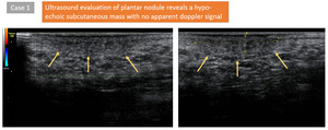
Fig. 1:
Case 1

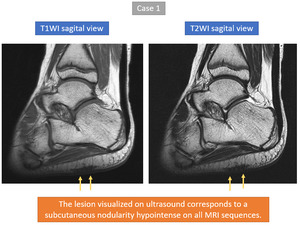
Fig. 2:
Case 1

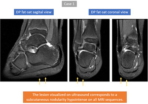
Fig. 3:
Case 1

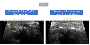
Fig. 4:
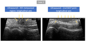
Case 2

Fig. 5:
Case 2

Fig. 6:
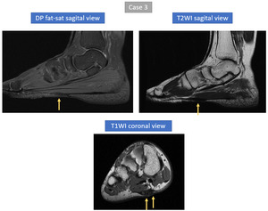
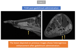
Case 3

Fig. 7:
Case 3

Fig. 9:
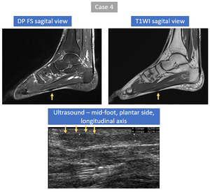
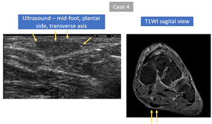
Case 4

Fig. 10:
Case 4

Fig. 11:
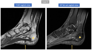
Case 5

Fig. 12:
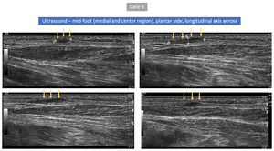
Case 6

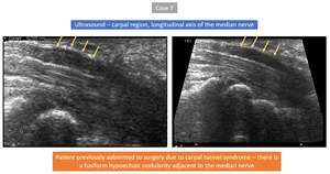
Fig. 13:
Case 7

Fig. 8:
Case 3