
ESSR 2019 / P-0054
anatomy and variants of intra-articular gleno-humeral structures (Biceps Brachii tendon, bicipitolabral complex, labrum and glenohumeral ligaments)
Congress:
ESSR 2019
Poster Number:
P-0054
Type:
Educational Poster
Keywords:
Musculoskeletal joint, Anatomy, MR, CT, Arthrography, Diagnostic procedure, Education and training
Authors:
I. Bougamra1, H. Riahi2, M. Chelli Bouaziz2, M. F. Ladeb2; 1Neuchâtel/CH, 2Tunis/TN
DOI:
10.26044/essr2019/P-0054
 in lateral surgical view.
LT (little tuberosity). GT (Greater tuberosity) References: Moser TP et al. Skeletal Rad 2015; 44:223-231")
Fig. 1:
supraspinatus aponeurotic expansion (SSp) in lateral surgical view.
LT (little...
 and coronal T2 with fat saturation AMR (b).")
Fig. 2:
supraspinatus aponeurotic expansion in axial DP with fat saturation (a) and...
")
Fig. 3:
Bicipito-labral complex type 1 with tight attachement to the glenoïd bone....

Fig. 4:
AMR : coronales T2 FS. BLC type 1
 in coronal anatomic view. References: 2019 Elsevier, Inc (Statdx)")
Fig. 5:
Bicipito-labral complex type 2 (thin sublabral recess) in coronal anatomic...
.
axial DP with fat saturation (a) and coronal T2 with fat saturation AMR (b)")
Fig. 6:
Bicipito-labral complex type 2 (thin sublabral recess).
axial DP with fat...
 in coronal anatomic view References: 2019 Elsevier, Inc (Statdx)")
Fig. 7:
Bicipito-labral complex type 3 (deep sublabral recess) in coronal anatomic view
.
axial DP with fat saturation (a) and coronal T2 with fat saturation AMR (b).")
Fig. 8:
Bicipito-labral complex type 3 (deep sublabral recess).
axial DP with fat...
")
Fig. 9:
sagittal view of glenohumeral ligaments
")
Fig. 10:
rotator cuff intervall
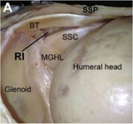
Fig. 11:
Rotator cuff interval (RI)
anatomy:
BT (biceps tendon), SSP (supra spinatus)

Fig. 12:
axial DPFS showing the biceps pouley. CHL: Coracohumeral ligament. SGHL :...
, T1 with fat saturation (b) and DP with fat saturation (c) showing the biceps pouley at the level of the coracoid. CHL: Coracohumeral ligament. SGHL : Superior glenohumeral ligament. LHB: long head of biceps tendon.")
Fig. 13:
sagittal T1(a), T1 with fat saturation (b) and DP with fat saturation (c)...
, T1 with fat saturation (b) and DP with fat saturation (c) showing the biceps pouley one slice lateral. CHL: Coracohumeral ligament. SGHL : Superior glenohumeral ligament. LHB: long head of biceps tendon.")
Fig. 14:
sagittal T1(a), T1 with fat saturation (b) and DP with fat saturation (c)...
, T1 with fat saturation (b) and DP with fat saturation (c) showing the biceps pouley. CHL: Coracohumeral ligament. SGHL : Superior glenohumeral ligament. MGHL : middle glenohumeral ligament. IGHL : Inferior glenohumeral ligament. LHB: long head of biceps tendon.")
Fig. 15:
sagittal T1(a), T1 with fat saturation (b) and DP with fat saturation (c)...
, T1 with fat saturation (b) and DP with fat saturation (c) showing the biceps pouley one slice lateral. CHL: Coracohumeral ligament. SGHL : Superior glenohumeral ligament. LHB: long head of biceps tendon.")
Fig. 16:
sagittal T1(a), T1 with fat saturation (b) and DP with fat saturation (c)...
, T1 with fat saturation (b) and DP with fat saturation (c) showing the biceps pouley one slice lateral. CHL: Coracohumeral ligament. SGHL : Superior glenohumeral ligament. LHB: long head of biceps tendon.")
Fig. 17:
Biceps pouley:
sagittal T1(a), T1 with fat saturation (b) and DP with fat...
, T1 with fat saturation (b) and DP with fat saturation (c) showing the biceps pouley one slice lateral. CHL: Coracohumeral ligament. SGHL : Superior glenohumeral ligament. LHB: long head of biceps tendon.")
Fig. 18:
Biceps pouley:
sagittal T1(a), T1 with fat saturation (b) and DP with fat...
")
Fig. 19:
rotator cable
 and T1 (b) showing the CHL (Coracohumeral ligament) and his posterior part called rotator cable.")
Fig. 20:
sagittal T1 with fat saturation (a) and T1 (b) showing the CHL (Coracohumeral...
 and axial DP with fat saturation (b) showing the CHL (Coracohumeral ligament) and his posterior part called rotator cable in a case of PASTA lesion.")
Fig. 21:
sagittal T1 with fat saturation (a) and axial DP with fat saturation (b)...
. Anatomy of the capsulolabral complex and rotator interval related to glenohumeral instability. Knee Surgery, Sports Traumatology, Arthroscopy, 24(2), 343–349.")
Fig. 22:
Labrum anatomy
 and a rounded appearance (b) References: Service de Radiologie Pr M Chelli Bouaziz. Institut MT Kassab d'orthopédie. Tunisie")
Fig. 23:
Normal glenoid labrum.
These images is at the junction of the middle and...
 and type B (b) chondrolabral junction")
Fig. 24:
axial DP fat saturation showing type A (a) and type B (b) chondrolabral...
")
Fig. 25:
sagittal view of normal labrum and glenohumeral ligaments

Fig. 26:
labral foramen
 with successive slices.")
Fig. 27:
sublabral foramen in sagittal DP Fat Saturation MRI ( a and b) with successive...

Fig. 28:
Labral foramen. Arthro-CT

Fig. 29:
Labral foramen. Arthro-MR

Fig. 30:
labrum absence

Fig. 31:
absence of labrum in sagittal DP Fat Saturation AMR
")
Fig. 32:
Buford complex. Labrum absence and thick MGHL
 and axial DP Fat Saturation MRI (b)
showing the absence of anterosuperior labrum (1) and a thick middle glenohumeral ligament (2).")
Fig. 33:
buford complex in sagittal T1 (a) and axial DP Fat Saturation MRI (b)...

Fig. 34:
Arthro-MR : association of labral recessus and foramen in axial DP FS and...

Fig. 35:
Arthro-MR : association of labral recessus and foramen in axial DP FS
 and without fat saturation (b) showing the SGHL (superior glenohumeral ligament), MGHL (middle glenohumeral ligament) and IGHL (Inferior glenohumeral ligament)")
Fig. 36:
sagittal T1 with fat saturation (a) and without fat saturation (b) showing the...
 underneath coracoid process.
References: Service de Radiologie Pr M Chelli Bouaziz. Institut MT Kassab d'orthopédie. Tunisie")
Fig. 37:
Oblique sagittal reformatted image of CT arthrography shows SGHL (arrow)...
 and MGHL (arrow) have a long common origin off the anterior labrum
References: Service de Radiologie Pr M Chelli Bouaziz. Institut MT Kassab d'orthopédie. Tunisie")
Fig. 38:
The SGHL (arrowhead) and MGHL (arrow) have a long common origin off the...
. Anatomy of the superior glenohumeral ligament. Journal of Shoulder and Elbow Surgery, 19(6), 908–916.")
Fig. 39:
MGHL anatomy
. Axial (a) and oblique sagittal (b) images. Note the straight appearance of the ligament
References: Service de Radiologie Pr M Chelli Bouaziz. Institut MT Kassab d'orthopédie. Tunisie")
Fig. 40:
Normal MGHL (arrow). Axial (a) and oblique sagittal (b) images. Note the...

Fig. 41:
double MGHL in sagittal T1 FS AMR on the left versus a normal anatomy on the...

Fig. 42:
Arthro-MR : sagittal and coronal T2 FS blade showing the IGHL
. Anatomy of the capsulolabral complex and rotator interval related to glenohumeral instability. Knee Surgery, Sports Traumatology, Arthroscopy, 24(2), 343–349.")
Fig. 43:
attachement types of IGHL
 in sagittal T1 with fat saturation (a) and T1 MRI (b) . IGHL (1), CHL (2) and SGHL (3).")
Fig. 44:
High attachement of the inferior glenohumeral ligament (IGHL) in sagittal T1 ...
 and Oblique sagittal (b) images show inferior glenohumeral ligament (long arrows) running parallel to the anterior inferior glenoid labrum.
References: Service de Radiologie Pr M Chelli Bouaziz. Institut MT Kassab d'orthopédie. Tunisie")
Fig. 45:
Arthro-CT : Axial (a) and Oblique sagittal (b) images show inferior...
 in axial DP with fat saturation going from up (a, b) to down (c, d) .")
Fig. 46:
High attachement of the inferior glenohumeral ligament (IGHL, arrow) in axial...