ESSR 2019 / P-0115
Migratory loose bodies from the ankle joint to the flexor hallucis longus tendon sheath – Imaging features
Congress:
ESSR 2019
Poster Number:
P-0115
Type:
Educational Poster
Keywords:
Education and training, Normal variants, Education, Ultrasound, MR, CT, Musculoskeletal soft tissue, Musculoskeletal joint
Authors:
J. S. Wong, V. Cassar-Pullicino; Oswestry/UK
DOI:
10.26044/essr2019/P-0115
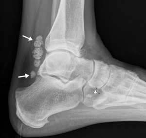
 and a zone 2 loose body (arrowhead).")
Fig. 1:
Radiograph demonstrating several zone 1 loose bodies (white arrows) and a zone...
 in image (a), which altered to a radiolucent appearance with a sclerotic rim in (b). Note slight increase in size.")
Fig. 2:
Radiographs taken 24 months apart, demonstrating initial dense, trabeculated...
, (b) and (c) demonstrating decrease in size of the zone 2 loose bodies. Note that the larger loose bodies in image (a) are orientated with their largest dimension lying longitudinally along the tendon sheath. There is severe osteoarthritis in the ankle joint and instability with anterior translation of the talus, prior to ankle fusion in image (c).")
Fig. 3:
Serial radiographs (a), (b) and (c) demonstrating decrease in size of the zone...
 T1 sagittal and (b) T1 TIRM sagittal images demonstrating a loose body with high marrow fat content in the FHL tendon sheath.")
Fig. 4:
(a) T1 sagittal and (b) T1 TIRM sagittal images demonstrating a loose body with...
 T1 and (b) T1 TIRM sagittal images. They are ossified on radiograph (c).")
Fig. 5:
Loose bodies in zone 2 of the FHL tendon sheath, demonstrated as low signal...
, demonstrated as a low signal structure on (a) sagittal T1 and (b) sagittal T1 TIRM images. It is unmineralised on CT, image (c). Note a co-existing ossified loose body immediately caudally (arrowheads).")
Fig. 6:
Cartilaginous loose body in zone 1 FHL tendon sheath (arrows), demonstrated as...
 T1 sagittal image demonstrating a loose body in the FHL tendon sheath (arrow) with high T1 signal. (b) It has some high signal on this PD FS axial image. (c) This loose body is not mineralised on CT. Appearance is in keeping with lipometaplasia of a cartilaginous loose body.")
Fig. 7:
(a) T1 sagittal image demonstrating a loose body in the FHL tendon sheath...
 zone 1 and (b) zone 2 of the FHL tendon sheath. Note the hyperechogenic reflectivity with dense posterior acoustic shadowing.")
Fig. 8:
Ultrasound images of two different patients, showing the FHL tendon sheath in...
 in zone 1 FHL tendon sheath, seen on the lateral radiograph (a), same patient as in Fig. 6. (b) Axial PD FS image demonstrating the loose body clearly within the FHL tendon sheath (arrow), and effacing the FHL muscle and tendon.")
Fig. 9:
Spiculated loose body (arrow) in zone 1 FHL tendon sheath, seen on the lateral...