Keywords:
Trauma, Diagnostic procedure, Ultrasound, MR, Musculoskeletal joint
Authors:
G. Kiernan, Z. Qamhawi, K. Shah, C. Azzopardi, J. Teh; Oxford/UK
DOI:
10.26044/essr2019/P-0147
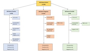
Imaging findings OR Procedure Details
Over a 5 year period 228 patients were identified using a key word search of the radiology information system (RIS).
The surgical notes were then reviewed from all those pateint who proceded to surgery.
All ultrasounds were conducted by MSK Consultant radiologist or Fellow.
191 were deemed to be negative for Stener lesion on US imaging,
of these 9 patients proceeded for surgical exploration on clinical grounds and all were found to be negative for Stener lesion.
28 were deemed to be positive for Stener lesion on US imaging however intraoperative finings deemed 8 positive and 8 negative.
9 were considered possible stener lesion on US,
3 of these preceded for surgery and all were negative.
Fig. 3: Flow chart of results comparing ultrasound findings to intraoperative findings.
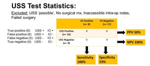
Fig. 4: Stastistical results displaying sensitivity, specificity, positive predictive value and neagtive predictive value.
In this cohort of pateints 14 proceeded to MRI of the thumb.
12 of these were fromt the US negative group and 2 from the US possible group.
On MRI all 12 of the US negative were also MRI negative for stener lesion.
Of the 2 US possible 1 was neagative and 1 was positive for stener lesion.
This MRI postive case was also positive for stener lesion intraoperatively.
Limitations:
The limitations of this study include a single centre retrospective study,
small sample size and low prevalence of stener lesion.