ESSR 2019 / P-0163
MDM2 in Atypical Lipomas
Congress:
ESSR 2019
Poster Number:
P-0163
Type:
Scientific Poster
Keywords:
Tissue characterisation, Neoplasia, Laboratory tests, Biopsy, MR, Musculoskeletal soft tissue
Authors:
S. Al-Qassab, P. Suresh, L. Farmkiss, K. Syred; Plymouth/UK
DOI:
10.26044/essr2019/P-0163

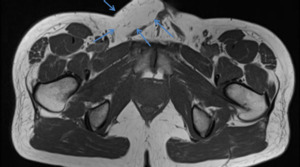
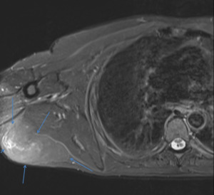
Fig. 1:
Axial T1 MRI image demonstrating a subcutaneous fatty lesion in the right groin.

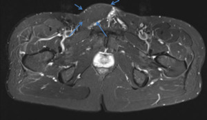
Fig. 2:
Axial STIR MRI image demonstrating complete suppression of the fatty lesion in...

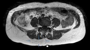
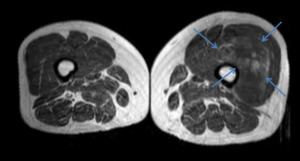
Fig. 3:
Axial T1 MRI image demonstrating a subcutaneous fatty lesion in the back right...

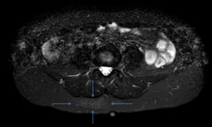
Fig. 4:
Axial STIR MRI image demonstrating subtle increased signal within the...

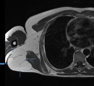
Fig. 5:
Axial T1 MRI image demonstrating a subcutaneous fatty lesion in the right chest...

Fig. 6:
Axial STIR MRI image demonstrating high signal intensity with some nodules and...

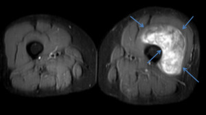
Fig. 7:
Axial T1 MRI image demonstrating a large heterogenous intramuscular lesion in...

Fig. 8:
Axial STIR MRI image demonstrating diffuse high signal intensity within the...