ESSR 2019 / P-0163
MDM2 in Atypical Lipomas
Congress:
ESSR 2019
Poster Number:
P-0163
Type:
Scientific Poster
Keywords:
Tissue characterisation, Neoplasia, Laboratory tests, Biopsy, MR, Musculoskeletal soft tissue
Authors:
S. Al-Qassab, P. Suresh, L. Farmkiss, K. Syred; Plymouth/UK
DOI:
10.26044/essr2019/P-0163

Fig. 9:
MDM2 results in the retroperitoneal lesions.

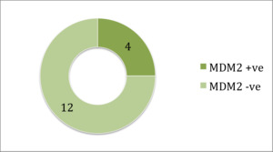
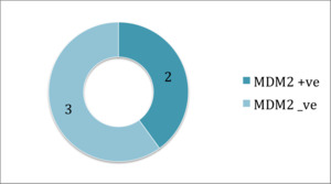
Fig. 10:
MDM2 positive in the non-retroperitoneal lesions.

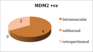
Fig. 11:
MDM2 positive cases distribution.

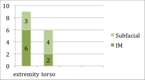
Fig. 12:
Anatomical distribution of the lesions.

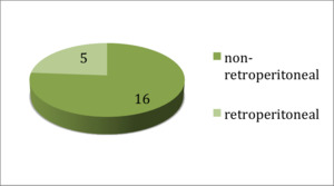
Fig. 13:
Distribution of retroperitoneal and non-retroperitoneal lesions.