
ECR 2018 / C-2480
Pleural pathology – a primer for radiologists
Congress:
ECR 2018
Poster Number:
C-2480
Type:
Educational Exhibit
Keywords:
Lung, Mediastinum, Respiratory system, CT, PET-CT, Digital radiography, Sampling, Diagnostic procedure, Biopsy, Pathology, Cancer, Infection
Authors:
A. Gangahar 1, Z. Wahab1, J. Zhong1, H. Nejadhamzeeigilani1, M. Kon2; 1Leeds/UK, 2Bradford/UK
DOI:
10.1594/ecr2018/C-2480

5. Are the costophrenic angles and hemidiaphragms well defined? (Any pleural effusion)")
Fig. 1:
Checklist of pleura and pleural space review on a frontal chest x-ray:
1....
.
Figure B demonstrates a larger right sided pleural effusion. On both x-rays the hemidiaphragm border of the affected side cannot be delineated.")
Fig. 2:
Chest x-rays showing pleural effusions. On figure A, there is a left sided...
 and lateral (B) chest x-rays demonstrated a loculated pleural effusion in the left oblique fissure.")
Fig. 3:
Frontal (A) and lateral (B) chest x-rays demonstrated a loculated pleural...
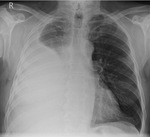
Fig. 4:
The patient, a non-smoker, presented with shortness of breath and cough for...