Purpose
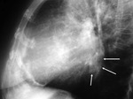
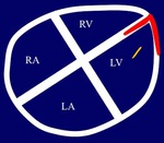
Describe uncommon and atypical cardiovascular calcifications.
Illustrate the imaging findings on chest radiograph and multislice CT.
Discuss the usefulness of these techniques for diagnosing cardiovascular calcifications,
including uncommon and atypical.
Methods and Materials
Uncommon and atypical cardiovascular calcifications are collected retrospectively from a teaching data base in our hospital.
Results
IMAGING TECNIQUES
CHEST RADIOGRAPH
Initial and sometimes only study to provide information regarding structure and function of the cardiovascular system.
Allows assessing size of cardiac chambers,
calcifications,
left and right cardiac dysfunction,
pulmonary circulation,
pulmonary artery hypertension and congenital heart abnormalities.
Calcifications are better depicted in an overexposed radiograph and with a tangential plane,
though superposition of other structures (i.e.
heart or spine) may hamper its visualization.
MULTISLICE CT
Very sensitive technique for detection,
localization and quantification of cardiovascular calcifications.
Particularly useful for coronary disease...
Conclusion
Cardiovascular calcifications,
including uncommon and atypical ones,
are associated with several causes and their study by chest radiograph and multislice CT is essential for the diagnosis.
References
Gowda RM,
Boxt LM.
Calcifications of the heart.
Radiol Clin N Am.
2004;42:603-17.
Ferguson EC,
Berkowitz EA.
Cardiac and pericardial calcifications on chest radiographs.
Clin Radiol.
2010;65(9):685-94.
Lee VS,
Patz EF,
Chen JTT.
Atypical and unsusual calcifications of the heart and great vessels: Imaging findings.
AJR Am J Roentgenol.
1994;163:1349-55.
Harpaz D,
Auerbach I,
Vered Z,
Motro M,
Tobar A,
Rosenblatt S.
Caseous calcification of the mitral annulus: a neglected,
unrecognized diagnosis.
J Am Soc Echocardiogr.
2001;14(8):825-31.
Araoz PA,
Eklund HE,
Welch JF.
CT and...


