Purpose
CT is a valuable technique for prosthetic heart valve (PHV) assessment
CT helps identify the cause of PHV dysfunction including obstructive masses (pannus/thrombus) and endocarditis (vegetations,
mycotic aneurysms).Retrospective ECG-gated CTA is most often used with reconstructions at each 5-10% of R-R interval which are needed to dynamically assess leaflet motion and anatomy.
Howver,
this results inhigh radiation exposure for the patient.
Furthermore,
the ideal scan protocol would include not only acontrast enhanced ECG-gated acquisition with reconstructions at each 5-10% R-R interval but also a non...
Methods and Materials
We retrospectively analyzed the CT scans of patients that were scanned according to the acquistion protocol described below.
The CT acquisition protocol consisted of 3 sequential acquisitions:
1.
Non contrast enhanced prospectively ECG-triggered scan of PHV only at 75% of the R-R interval.
2.Wide pulse window sequential CTA with recons at each 5%,
iterative reconstruction ADMIRE level 4 (Figure 1).For the prospectively ECG-gated sequential scan3 stacks were planned with the middle stack centered on the valve.
The maximum scanlength for 3 stacks in the z-axis...
Results
Scans of 10 patients with 11 prosthetic heart valves were found and analyzed.
Image quality scores were:
Non contrast enhanced scan: 4.0±1.8*
Contrast enhanced scan: 4.8±0.4
Venous phase scan: 4.2±0.4
Values as mean ± SD; 1=non-interpretable; 5= excellent,
*n=9
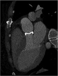
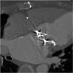
Figures 2 and 3 illustrate different scores of image quality.
CTDI values and radiation dose were:
Non contrast enhanced scan: 3.33±1.91 mGy
Contrast enhanced scan: 32.45±15.19 mGy
Venous phase scan: 2.53±1.51mGy
Total dose : 8.4±3.8mSv
Values as mean±SD; total dose for n=9,
dose info not available...
Conclusion
Comprehensive assessment of prosthetic heart valves at moderate radiation dose is possible using dual source CTA an iterative reconstruction.


