Keywords:
Imaging sequences, CT-Angiography, CT, Cardiac, Prostheses
Authors:
R. P. J. Budde, M. L. Dijkshoorn, J. Bekkers, L. E. Swart, M. van Straten, K. Nieman, G. P. Krestin; Rotterdam/NL
Results
Scans of 10 patients with 11 prosthetic heart valves were found and analyzed.
Image quality scores were:
Non contrast enhanced scan: 4.0±1.8*
Contrast enhanced scan: 4.8±0.4
Venous phase scan: 4.2±0.4
Values as mean ± SD; 1=non-interpretable; 5= excellent,
*n=9
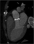
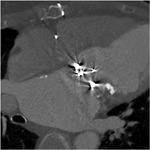
Figures 2 and 3 illustrate different scores of image quality.
CTDI values and radiation dose were:
Non contrast enhanced scan: 3.33±1.91 mGy
Contrast enhanced scan: 32.45±15.19 mGy
Venous phase scan: 2.53±1.51mGy
Total dose : 8.4±3.8mSv
Values as mean±SD; total dose for n=9,
dose info not available in 1 patient, abdomen included in venous phase scan in one patient.
Dynamics of mechanical valve leaflet motion could be succesfully assessed (figure 4). PHV findings included: normal function (n=6),
obstruction,
leakage or aortic root cavities (n=4),
non-assessable (n=1).
The value of the non-contrast enhanced and delayed phase scans are illustrated in figures 5 to 7.


