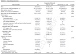
1. Patient characteristics (Fig.
6)
◦ Ring type : 27 CE ring vs.
18 Duran ring
◦ Significant difference in mean time after MV annuloplasty between ring types (P<0.05)
2.
CT Analysis of Annuloplasty Rings
◦ Pannus around the annuloplasty ring : 29 of 45 cases (64.4%)
◦ Significant pannus: 10 patients (22.2%)
◦ Leaflet thickening (>2mm): 31 of 45 patients (68.9%)
◦ Valve opening area : 2.34 ± 0.717 cm2 (n=22)
◦ Patients with significant pannus vs.
patients with no or insignificant
- Significant difference in valve opening area (1.71 cm2 vs 2.63 cm2; P<.05)
- No significant difference in valve leaflet thickenss (2.43 ± 1.11 mm vs 2.12 ± 0.794 mm ; P=.352)
3. Correlation Between CT Findings and TTE Parameters
◦The valve opening area on CT was positively correlated with MVA on TTE and negatively correlated with the mean diastolic PG (P<.05) (Fig.
7A-B)
◦With increasing pannus severity,
the mean diastolic PG was significantly elevated (Fig.
7C)

Fig. 7: Correlation between CT measurements and TTE parameters. Scattered diagram showing (a) a negative correlation between the mean PG on TTE and the maximal opening area on CT, and (b) a positive correlation between MVA on TTE and the maximal opening area measured on CT. Box and Whisker plots show differences in (c) the mean PG according to pannus severity.
◦ Thirteen patients (4 with CE rings and 9 with Duran rings) with functional MS(mean diastolic PG ≥ 5 mm Hg)
◦ Patients with functional MS vs.
patients with normal range PG
- A higher incidence of pannus and significant pannus in functional MS
: Pannus: 100 % vs.
50%; significant pannus: 69.2% vs.
3.13% (P<0.05)
: Even after adjustment of the interval between MV repair and CT scan
- Leaflet thickening and maximal opening area on CT were not different
4.
Comparison of CT and TTE Findings Between Duran and CE Ring (Fig.
8)
◦ TTE
- Patients with the Duran ring had a higher mean diastolic PG,
a smaller MVA,
and a higher incidence of functional MS than patients with the CE ring (P <.05)
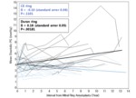
- PG was gradually elevated in the Duran ring group as time passed (0.19 mm Hg/y) to a statistically significant extent (P =.0018) but not in the CE ring group (Fig.
9)
◦ CT
- The proportion of pannus and significant pannus: Duran ring > CE ring
(even after adjustment for interval (P<.05))
- The mitral opening area on CT: Duran ring < CE ring (P<.05)
- The size of the inserted annuloplasty ring and the annular area measured on CT: not different
- Annular sphericity index: During ring <CE ring (P<.05)
: Duran ring is more circular
→ Difference in annular shape may cause more frequent development of pannus formation in the Duran ring
5.
Clinical course
◦ Among the entire study population of 45 patients,
3 patients underwent reoperation,
1 for MS after having the Duran ring inserted and another 2 for detachment of the Duran ring (Fig.
10-12).

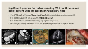
Fig. 10: Significant pannus formation causing MS in a 52-year-old male patient with the Duran annuloplasty ring.
CT image of a patient with a 31mm Duran ring shows leaflet thickening of the mitral valve (1st column) and severe pannus formation around the annular ring (2nd and 3rd columns). The patient underwent mitral ring annuloplasty 16 years before CT scanning because of subacute infective endocarditis. Mean-diastolic pressure gradient on TTE was elevated (18mmHg)(4th column). Severe fibrosis of mitral valve with a circumferential pannus was seen during redo-surgery (5th column).


