Patients:
• 31 consecutive patients (22M, 9F)
• Aged 47-73 ys: mean 61ys
• 1.5 tesla scanner with single shot EPI.
Methods:
• Routine MR (T1,T2WI) (n=31).
• DSC perfusion MRI (n=31).
• Routine post contrast study (n=31).
Data Acquisition
MR Machine:
• 1.5 tesla MR unit (symphony-Siemens).
• Gradient strength: 30mTm.
• slew rate: 120 T/m/s.
Single Shot DSC T2*WI:
• TR:120 ms, TE:47ms.
• NEX:1, Slice thick: 5mm.
• Slice no: 15, FOV: 20-30 cm.
Contrast medium injection
CM: Type: Gd-DTPA.
• Dose: 0.2 mmol/ kg BW.
• Rate: 5ml/s followed by 20ml saline.
• Time between data points: 2 sec.
• Total acquisition time: 2min.
Image analysis
• Quantitative assessment of DSC%
• ROI was placed along the margin of the tumor.
• Time signal intensity curve (TIC) was obtained.
• DSC%= unenhanced lesion SI (S0) -contrast enhanced lesion(SI)/unenhanced lesion SI (S0)
•
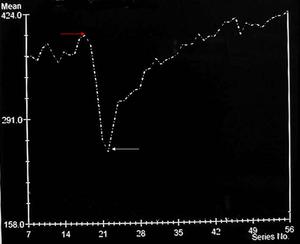
Fig.: DSC curve: Red arrow repesetn SO and white arrow represetn S1
Unenhanced SI: red arrow, enhanced SI white arrow.
Quantitative assessment of tumor volume:
• Multiple section at post contrast MR study.
Final diagnosis
Pathological examination:
• Tru cut biopsy were done for all patients during fibro-optic endoscopy.
Pathological type:
• According to WHO classification Type I, II, II.
Pathological grade:
• well, moderately, poorly and undifferentiated.
Statistical analysis
Mean & SD:
T test (P value):
Pearsons Correlation:
- DSC% correlated with 4 prognostic parameters.

