Type:
Educational Exhibit
Keywords:
Neuroradiology brain, Neuroradiology peripheral nerve, Neuroradiology spine
Authors:
R. Calandrelli, S. Gaudino, G. Di Lella, T. Tartaglione, A. M. Costantini, A. Pedicelli, C. Colosimo; Rome/IT
DOI:
10.1594/ecr2010/C-2698
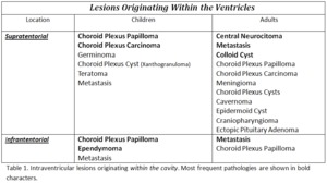
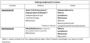
Background
A lesion may be considered intraventricular if it is found primarily within the ventricular system, causing expansion of the ventricle; therefore, it may be sometimes difficult to establish the exact origin of a lesion, for example to assess if a mass extends from the ventricular wall to the ventricular cavity or vice-versa.
Nevertheless, we may classify ventricular lesions in two main categories: those that originate from the ventricular walls (i.e. from the subependymal / subventricular region) and subsequently grow into the ventricle, and those that originate from structures within the ventricular system, such as the choroid plexus. (Fig. 1)
The differential diagnosis list may be addressed by integrating clinical data (e.g. Patients’ age, some inherited specific syndromes), together with morphological (CT/MRI) and some “non-morphological” RM imaging findings (DWI, MRS).
The most common pathologies are listed in the tables below, according by preferential site of origin and age.
![](https://epos.myesr.org/posterimage/esr/ecr2010/102688/media/298654?maxheight=300&maxwidth=300)
Fig.
![](https://epos.myesr.org/posterimage/esr/ecr2010/102688/media/298655?maxheight=300&maxwidth=300)
Fig.