ECR 2010 / C-2713
Radiology of hypothalamic lesions: A pictorial essay depicting characteristic hypothalamic pathologies
Congress:
ECR 2010
Poster Number:
C-2713
Type:
Scientific Exhibit
Keywords:
Neuroradiology brain, Neuroradiology peripheral nerve, Neuroradiology spine
Authors:
A. J. B. Baxi, M. belman, T. Nagendra, S. Vidyasagar, K. L. Tourani; Hyderabad/IN
DOI:
10.1594/ecr2010/C-2713
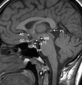
.T1 sagittal demonstrates anatomy of hypothalamus.
AC-anterior commissure,PC-posterior commissure,MB-mamillary bodies,
TC-tuber cinerium,IS-infundibular stalk,OC-optic chiasma,PP-posterior pituitary gland.")
Fig. 1:
Fig(1).T1 sagittal demonstrates anatomy of hypothalamus.
AC-anterior...
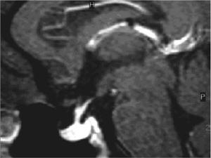
.Sagittal T1 contrast- the infundibular stalk and pituitary gland show normal homogeneous enhancement, which reflects their lack of a blood-brain barrier.")
Fig. 2:
Fig(2).Sagittal T1 contrast- the infundibular stalk and pituitary gland show...
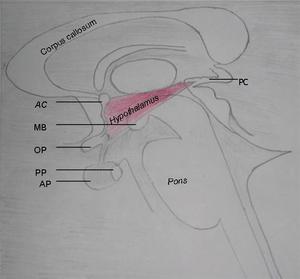
AC-Anterior commissure,PC-Posterior commissure,MB-Mamillary bodies,PP-Posterior pituitary,AP-Anterior pituitary.")
Fig. 3:
Fig1.Drawing shows the hypothalamus (outlined with a pink color)AC-Anterior...