Keywords:
Neuroradiology brain, Neuroradiology peripheral nerve, Neuroradiology spine
Authors:
A. J. B. Baxi, M. belman, T. Nagendra, S. Vidyasagar, K. L. Tourani; Hyderabad/IN
DOI:
10.1594/ecr2010/C-2713
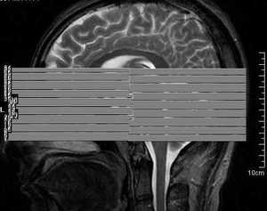
Methods and Materials
A retrospective review of our institutional case archive from 2002 to 2009 revealed broad range of neoplasms, inflammatory and infectious disorders, and congenital and developmental lesions. The region of study included hypothalamus, pituitary gland and parasellar region.
Imaging were performed on 1.5 Tesla MRI with head coil, single slice CT, 6 slice CT and 64 slice CT scanner to characterize the pathologic spectrum of hypothalamic and parahypothalamic diseases with soft tissue and bony reconstruction whenever needed.
Sagittal and coronal spin-echo T1-weighted sequences were performed with thin sections (3mm) and a small field of view (200-230mm). The axial sections were set at 4 mm thickness. The same sequence was repeated after the intravenous administration of a standard dose (0.2mmol/kg) of gadopentetate dimeglumine and 1 mm spacing.(Fig1-3)
![](https://epos.myesr.org/posterimage/esr/ecr2010/102703/media/298997?maxheight=300&maxwidth=300)
Fig.: Sagittal localiserfor planning axial images
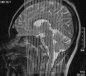
![](https://epos.myesr.org/posterimage/esr/ecr2010/102703/media/298999?maxheight=300&maxwidth=300)
Fig.: Localiser for coronal planning
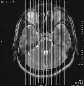
![](https://epos.myesr.org/posterimage/esr/ecr2010/102703/media/299001?maxheight=300&maxwidth=300)
Fig.: Localizer for coronal planning
All patients had sagittal T1, T2 weighted, axial T1, T2 weighted with fat saturation, Flair, coronal T1, Flair, and post-gadolinium axial, sagittal and coronal T1 weighted sequences. In addition, in some patients, sagittal dynamic image were also done if small pituitary and hypothalamic lesions were suspected. This dynamic study was done after rapid injection of gadopentetate dimeglumine.