ECR 2010 / C-3262
Digital Breast Tomosynthesis (DBT): A Useful Adjunct To Two Dimensional Mammography (2DM) For The Preoperative Localization Of Subtle Soft Tissue Lesions
Congress:
ECR 2010
Poster Number:
C-3262
Type:
Educational Exhibit
Keywords:
Breast
Authors:
S. Mombelloni, A. Iqbal, D. R. Evans, R. K. Wasan, J. C. Morel, C. Peacock, M. J. Michell; London/UK
DOI:
10.1594/ecr2010/C-3262

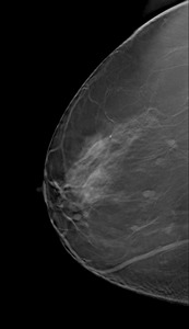
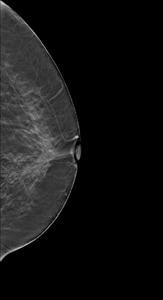
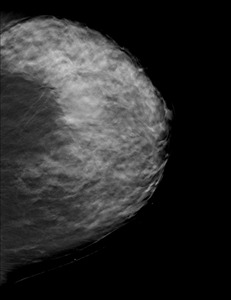
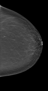
Fig. 1:
FIRST CASE: DBT

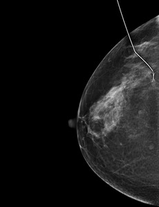
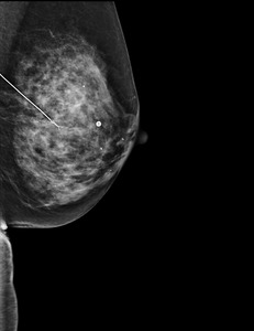
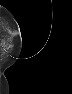
Fig. 2:
2D post wire

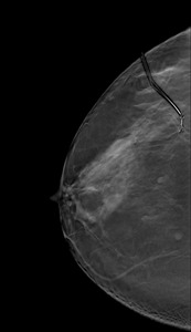
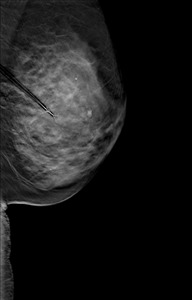
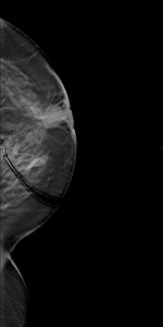
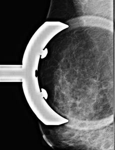
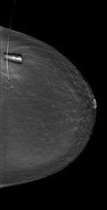
Fig. 3:
DBT post wire

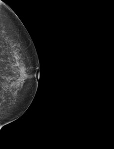
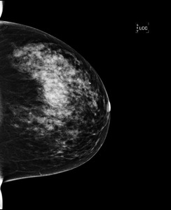
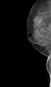
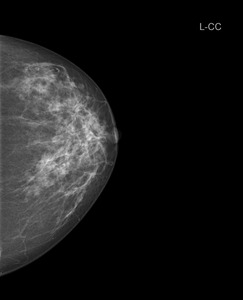
Fig. 4:
SECOND CASE: 2D Mag

Fig. 5:
2D post wire

Fig. 6:
DBT post wire

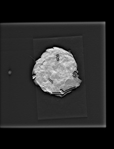
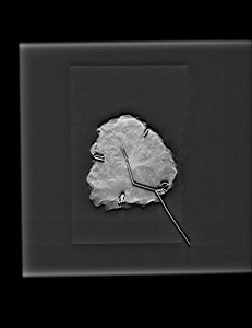
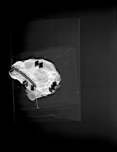
Fig. 7:
DBT specimen

Fig. 8:
THIRD CASE: 2D

Fig. 9:
DBT

Fig. 10:
2D post wire

Fig. 11:
DBT post wire

Fig. 12:
FOURTH CASE: 2D

Fig. 13:
DBT

Fig. 14:
DBT post wire

Fig. 15:
FIFTH CASE 2D Mag

Fig. 16:
DBT post wire

Fig. 17:
DBT specimen

Fig. 18:
SIXTH CASE: DBT

Fig. 19:
DBT post wire

Fig. 20:
SEVENTH CASE: 2D

Fig. 21:
DBT specimen