Keywords:
Motility, Diverticula, Dilatation, Technical aspects, Barium meal, Barium enema, Teleradiology, Conventional radiography, Education and training, Animal (veterinary) studies
Authors:
V. vulpe1, C.-A. Vulpe1, L. Paval2; 1Iasi/RO, 2Pascani/RO
DOI:
10.1594/ecr2011/C-1264
Results
Our studies have shown that the barium sulphate solution that was administred to the pigeons (found in a light estate of stress,
because of the handling) passes too quickly through the glandular and the mechanic stomach,
which cause this area to remain unmarked,
from a radiologic point of view.
Thus,
we thought of delaying the peristaltism using an anesthetic – in one of the cases,
we administred Ketamine in a dose of 2 mg/100 g of body weight,
after which we proceeded with the radiologic examination,
in a latero-lateral position (the pigeon was held on its’ right side,
with the wings fastened to the back,
and the legs drawn to the back).
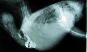
Fig.: Lateral radiographic image. The presence of the substance of contrast inside the ingluvia.
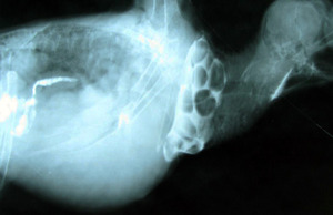
Fig.: The rarefaction of the sollution as it enters the intestine.
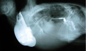
Fig.: Lateral radiographic image; the dillatation of the ingluvia, the penetration of the solution into the mechanical stomach and its’ diffusion inside the intestine.
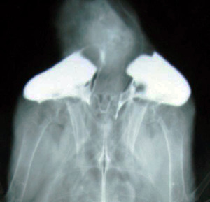
Fig.: Dorso-ventral image, with the formation of 2 ingluvial sacks.
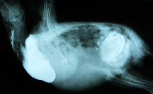
Fig.: Lateral radiographic image – the presence of the solution of contrast inside the intestine and in the terminal portion of the intestine.
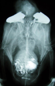
Fig.: Dorso-ventral radiographic image – the two ingluvial sacks and the substance of contrast that has reached the cloaca.
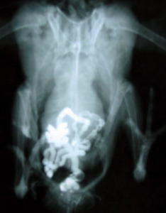
Fig.: Dorso-ventral image, showing the presence of the solution inside the cloaca and the terminal portion of the intestine.
