Clinical data
29 patients
- M : F = 11 : 18
- mean age : 68 years (range 39~92 years)
- Symptoms : a sudden lower abdominal pain in all patients
- Past medical history
- chronic debilitating underlying diseases : 12 patients (41.4%) - such as DM,
CRF,
CHF,
and Liver cirrhosis
- constipation : 4 patients (13.8%)
- Past GI tract operation history : 5 patients (17.2%)
Pathologic examination
- Confirmed as non-specific chronic or acute inflammation with colonic wall perforation
- Accompanying necrosis,
acute serositis,
diffuse hemorrhage and vascular congestion with abundant neutrophil infiltration in the surrounding colonic mucosa
- Accompanying stercoral colitis caused by fecal impaction in 3 cases
Fig. 1: 1A. Perforation of the bowel wall (HE, x 40)
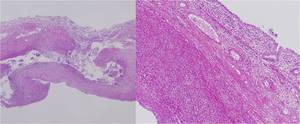
1B. Ascending colonic wall thining, acute and chronic inflammatory infiltrates with fibrinoid vasculitis, and ischemic change in the whole layer of the ascending colon wall (HE, x 100)
CT findings
- The most common perforation site: sigmoid colon (60%,
18/30)
- Other perforation site
ascending colon (10%,
3/30)
transvere colon (10%,
3/30)
descending colon (13.3%,
4/30)
colostomy site (6.7%,
2/30)
- Specific findings of colonic perforation
focal colonic wall defects (76.6%,
23/30)
fecal spillage (70%,
21/30)
extraluminal free air (100%,
30/30)
--> All cases had two of them at least.
Fig. 2: 2A. 70-year-old woman with spontaneous sigmoid colon perforation. Coronal image of three phase abdominal CT (venous phase) 
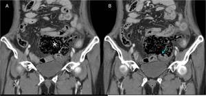
shows large amount of fecal spillage(*) from distal sigmoid colon and continuous sigmoid colonic wall (arrow).
2B. There is sigmoid colonic wall defect (blue arrow) on the next coronal CT scan image.
Fig. 3: 3A. Abdominal CT shows large amount of fecal spillage in pelvic cavity(*).
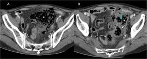
3B. On the 2.5 cm inferior from Figure 3A, there is focal sigmoid colonic wall defect (blue arrow) and fecal spillage from the defect site(*). There is diffuse edematous colonic wall thickening of sigmoid colon as a secondary sign of peritonitis(white arrows).
Fig. 4: 71-year-old woman with spontaneous colostomy site perforation.
Contrast enhanced abdominal CT shows extraluminal fee air(*) and fecal spillage(arrow) from perforation site of colostomy.
- Secondary peritonitis signs
pericolic fat infiltration (96.7%,
29/30)
peritoneal thickening with enhancement (83.3%,
25/30)
fluid collection (93.3%,
28/30)
edematous colonic wall thickening (46.7%,
14/30)
Fig. 5: 72-year-old woman with spontaneous descending colon perforation.
On axial image and coronal image of abdominal CT show fluid collection(*) 
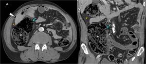
and colonic wall thickening(arrow).
There is extraluminal free air(*) as specific finding of colonic perforation.
Fig. 6: 74-year-old man with spontaneous ascending colon perforation with severe fecal impaction(*)
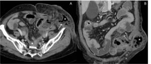
due to constipation.
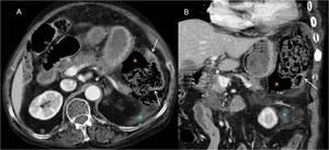
On axial image and coronal image of CT show fat infiltration(*), much dilatation of ascending colon up to 9 cm without any obstruction of colon. Some part of colonic wall is very thin(blue arrow). White arrow indicates ileocecal valve which is intact. Terminal ileum is normal.
There is extraluminal free air(arrowhead) as specific finding of colonic perforation.
Fig. 7: 7A. 50-year-old man with transverse colon perforation.
Plain abdomen erect radiograph shows pneumoperitoneum(*)
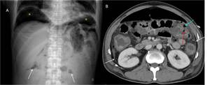
and air-fluid level without much dilatation of small bowel (arrow).
7B. Contrast enhanced CT shows peritoneal thickening with enhancement(arrows) as the sign of secondary peritonitis.
There is linear transverse colonic wall defect(red arrow) with free air just next to colonic wall defect.
CT findings
|
Sigmoid colon (18)
|
Ascending colon (3)
|
Transverse colon (3)
|
Descending colon (4)
|
Colostomy site (2)
|
Sum (30)
|
Colonic focal
wall defects
|
66.7% (12)
|
100% (3)
|
100% (3)
|
75% (3)
|
100% (2)
|
76.6% (23)
|
Fecal spillage
|
83.3% (15)
|
33.3% (1)
|
0% (0)
|
75% (3)
|
100% (2)
|
70.0% (21)
|
Extraluminal
free air
|
100% (18)
|
100% (3)
|
100% (3)
|
100% (4)
|
100% (2)
|
100% (30)
|
Edematous colonic wall thickening
|
50% (9)
|
0% (0)
|
100% (3)
|
25% (1)
|
50% (1)
|
46.7% (14)
|
Pericolic
fat infiltration
|
94.4% (17)
|
100% (3)
|
100% (3)
|
100% (4)
|
100% (2)
|
96.7% (29)
|
Fluid collection
|
94.4% (17)
|
66.7% (2)
|
100% (3)
|
100% (4)
|
100% (2)
|
93.3% (28)
|
Peritoneal thickening with
enhancement
|
88.9% (16)
|
66.7% (2)
|
66.7% (2)
|
100% (4)
|
50% (1)
|
83.3% (25)
|
Three patients died after surgery (10.3%,
3/29).
1) Two patients died within 3 days after surgery.
- One had two spontaneous colonic perforation.
--> In 2nd operation,
there was colostomy necrosis
and perfortation due to fecal impaction.
- One had bowel ischemia.
2) One patient died one month after surgery.
- The patient had peritonitis due to small bowel,
sigmoid colon and descending colon necrosis
one month after colon perforation.
- She had 2nd operation of total colectomy,
small bowel resection and anastomosis,
loop ileostomy and segmental resection of
sigmoid colon.
- Three days after 2nd surgery,
she had
another operation due to peritonitis
caused by ileostomy site perforation.