This poster was previously presented in Spanish at the 2010 Congreso Nacional SERAM (A Coruña)
Type:
Educational Exhibit
Keywords:
Neuroradiology brain, Vascular, CT-Angiography, CT, Contrast agent-intravenous, Image compression, Cerebrospinal fluid
Authors:
B. Prieto Hernández1, D. Santiago Agueda del Bas2, E. Gálvez González1, N. Alegre Borge1, N. D. Menocal Funez1, M. Villanueva Delgado1; 1Salamanca/ES, 2Avila/ES
DOI:
10.1594/ecr2012/C-0998
Background
Brain death marks an irreversible lack of brain functions.
In some dificult and complex cases of patients with metabolic disorders or patients treated with sedatives drugs,
in which the diagnosis of brain death after clinical examination,
additional test and EEG is subject to controversy, a brain CT with and without intravenous contrast can be helpful.
Our technique is based on two steps:
1- Brain CT scan without intravenous contrast.
2- CT angiography of the Willis Circle:
- We have to administered 100 cc of intravenous contrast at a rate of 3 cc/s.
- We have to make the sequences at 20 and 60 seconds after the administration of intravenous contrast.
The findings of brain death that we can check in the CT angiography are:
- Absence of intracranial flow visualization , that depends on the internal carotid artery and its branches.
- Persistent circulation in extracranial sistem,
that depends on the external carotid artery and its branches.
- Absence of flow visualization in vertebro-basilar system.
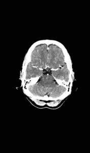
Fig. 2: Normal find CT angiography
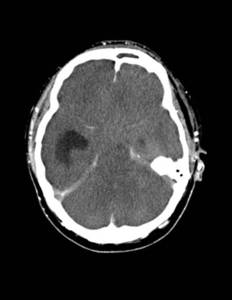
Fig. 1: No flow in intracranial arteries
Persistent circulation in the branches of the external carotid artery
