
ECR 2013 / C-1065
Can inflammatory myofibroblastic tumor (IMT) of the hepatobiliary system be differentiated from cholangiocarcinoma (CC) on imaging?
Congress:
ECR 2013
Poster Number:
C-1065
Type:
Scientific Exhibit
Keywords:
Abdomen, Liver, Oncology, CT, MR, Diagnostic procedure, Inflammation, Neoplasia
Authors:
R. Elias, F. Willemssen, K. Biermann, G. P. Krestin, R. Dwarkasing; Rotterdam/NL
DOI:
10.1594/ecr2013/C-1065

Fig. 1:
Delayed enhancement of IMT lesion.
No difference in enhancement pattern...

Fig. 2:
Delayed enhancement of CC lesion.
No difference in enhancement pattern between...

Fig. 3:
Multiple lesion in patient with IMT

Fig. 4:
Large, solitary lesion in patient with CC.
Notice capsule retraction

Fig. 5:
No bile duct dilatation in patient with IMT

Fig. 8:
Bile duct dilatation and vascular encasement in patient with CC

Fig. 7:
Large IMT lesion in segment IV of liver. Notice,no encasement of vena porta

Fig. 6:
Central CC lesion with bile duct dilatation and vascular encasement
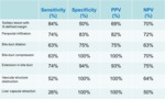
Fig. 9:
# Logistic regression model
# Tendency toward significance