ECR 2013 / C-1203
Diagnostic Value of Magnetic Resonance Imaging on Dermatofibrosarcoma Protuberans
Congress:
ECR 2013
Poster Number:
C-1203
Type:
Scientific Exhibit
Keywords:
Musculoskeletal soft tissue, Oncology, MR, Diagnostic procedure, Neoplasia
Authors:
J. Zhang, L. Ruan, S. Xu, B. QIAN; HANGZHOU/CN
DOI:
10.1594/ecr2013/C-1203

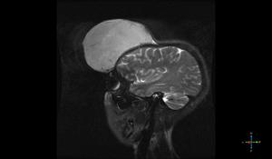
Fig. 1:
A 26-year-old female with recurrent DFSP of the frontal part.
Fig. 1:...

Fig. 3:
Fig.3: Axial T1WI shows a large mass of uniform iso-signal.

Fig. 4:
Fig.4: Axial T1WI with intravenous gadolinium administration shows apparent...

Fig. 6:
Fig. 6: Axial CT image of bone window shows no obvious bone destruction.
.")
Fig. 7:
Fig. 7: Photomicrograph shows monomorphous spindle-shaped cells arranged in a...

Fig. 2:
Fig. 2: Axial T2WI with fat suppression shows a large mass of uniform high...

Fig. 5:
Fig. 5: Axial CT image of brain window shows a soft-tissue mass similar to the...