ECR 2013 / C-1377
Performance of trained radiographers in lung nodule detection: prospective comparison with experienced radiologists in lung cancer screening
Congress:
ECR 2013
Poster Number:
C-1377
Type:
Scientific Exhibit
Keywords:
Thorax, Oncology, CT, CT-High Resolution, Screening
Authors:
A. Nair1, N. J. Screaton2, J. A. Holemans3, D. Jones3, L. Clements2, D. R. Baldwin4, J. K. Field3, D. M. Hansell1, A. Devaraj1; 1London/UK, 2Cambridge/UK, 3Liverpool/UK, 4Nottingham/UK
DOI:
10.1594/ecr2013/C-1377

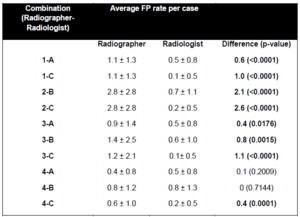
Fig. 1:
Frequency distribution of the number of nodules per CT study.

Fig. 2:
Breakdown of reference nodules according to category and nodule type. Numbers...
and C(n=130).")
Fig. 3:
Sensitivity of radiographer 1 compared to Radiologists A(n=130)and C(n=130).
and C(n=139).")
Fig. 4:
Sensitivity of radiographer 2 compared to Radiologists B(n=139)and C(n=139).
,B(n=87)and C(n=155).")
Fig. 5:
Sensitivity of radiographer 3 compared to Radiologists A (n=68),B(n=87)and...
,B(n=49)and C(n=113).")
Fig. 6:
Sensitivity of radiographer 4 compared to Radiologists A (n=64),B(n=49)and...