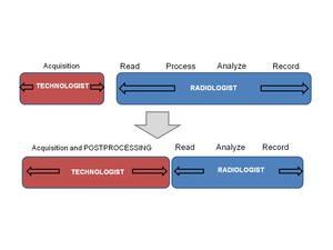
Type:
Educational Exhibit
Keywords:
Education and training, Computer Applications-General, MR-Diffusion/Perfusion, MR, CT, Computer applications
Authors:
C. Fraga Piñeiro, M. Centeno, M. González Vázquez, D. Castellón Plaza, G. Tardáguila, F. M. Tardaguila; Vigo/ES
DOI:
10.1594/ecr2013/C-1628
Imaging findings OR Procedure details
Postprocessing of CT and MRI images has proven to be a valuable tool in a variety of clinical applications due to its ability to provide additional diagnostic information.
It has applications in various organs and systems (neuroradiology,
musculoskeletal,
cardiovascular,
chest,
abdomen...).
It is essential to have image excellent quality original data to performance a clinical useful post-processing image.
Posprocessing techniques allow us to obtain anatomical information from the original images and also give us functional and molecular information.
We describe technical aspects and clinical applications of image post-processing techniques and we review from basic concepts to more advanced techniques.
1.
ANATOMICAL RECONSTRUCTIONS:
1.1 Basic techniques:
- MPR (Multi Planar Reformation): is a technique to show other planes that were not acquired directly during the acquisition.
This include sagittal,
oblique and coronal cross-sections reconstructed from axial images.
Fig. 1,
Fig. 2 .
Also it is possible to obtain curved plane to do so it is necessary to program a serie of points defining a curved path,
along which curved planes are reconstructed.
Reconstructions parallel to any anatomical structure could be obtained and is very useful for analysis of curved structures such as vessels or mandible. (Curved MPR) Fig. 3
- Subtraction: Subtraction imaging in MRI is a technique whereby an unenhanced T1-weighted sequence is digitally subtracted from the identical sequence performed after gadolinium administration.
By performing this operation,
any native T1 signal is removed and the remaining signal on the subtracted images is due solely to enhancement .It is very useful in breast MRI. Fig. 4
1.2 Advanced techniques:
- MIP( Maximum Intensity Projection):Consists on projecting the voxel with the highest attenuation value on every view throughout the volume on to a 2D image. This technique is very useful in CT or MRI angiography to show vessels enhanced with contrast.
MIP can have some artefacts,
for example: high-intensity material such as calcification can obscure information from intravascular contrast material and the use of the highest intensity value also increases the mean background intensity of the images and noise in the image.
Fig. 5 ,Fig. 6 ,Fig. 7
- miniMIP (Minimum Intesity Projection):Consists on projecting the voxel with the lowest attenuation value on every view throughout the volume on to a 2D image. It is particularly useful to visualize the biliary tract and pancreatic duct,
also to demonstrate organs filled air in CT.Fig. 8 ;Fig. 9
- AIP (Average Intensity Projection): The image represents the average of each component attenuation value.This can be useful for characterizing the internal structures of a solid organ or the walls of hollow structures such as blood vessels or intestine.
Fig. 10 . A variant of AIP is the "ray sum" in which each the attenuation value of each voxel is summed along the projection line. Therefore,
full-volume ray sum images may have an appearance similar to that of a conventional radiograph.
- VIP (Volumen Intensity Projection): assigns the highest attenuation values to the voxels that are closer to the viewer and lower values to those that are farther away,
thereby providing perspective information.
1.3 3D RECONSTRUCTIONS:
- SSD (Shaded Surface Display): In surface rendering,
the voxels located on the edge of a structure are identified,
usually by intensity thresholding,
and possibly enhanced with morphologic filtering or connectivity,
and these voxels are displayed.
The remaining voxels in the image are usually invisible.
This method actually shows only the surface of the organs as an opaque object.
Slicing through a surfaced rendered object will not reveal internal objects. It uses segmentation techniques or delimitation of the objects in the image,
ranging from pure drawing or contouring of a given structure manually to advanced algorithms (automatic segmentation).
This approach is useful for examining tubular structures,
such as the inside surfaces of airways (virtual bronchoscopy),
the colon (virtual colonoscopy),
and blood vessels.
But,
nowadays,
3D SSD is almost obsolete since it requires rather extensive work from the user and was replaced by volume rendering 3D reconstruction.Fig. 11
- VRD (volume-rendering): In this case not just the surface draw of the objects,
but also the inside,
adjusting the levels of transparency. In volume rendering,
the CT numbers that make up the image are assigned to be either visible or invisible,
to be displayed with varying colors,
and often to be displayed with varying opacity levels (transparency). It is widely used as part of CT and MRI,
and in various applications such as cardiovascular imaging, musculoskeletal applications and others.Fig. 12 ,
Fig. 13 .
2.
FUNCTIONAL TECHNIQUES:
- Diffusion and ADC ( Apparent diffusion coefficient): It is a technique based on random movement or Brownian movement of water molecules.
This Brownian motion is analogous to free diffusion,
however,
the water molecules movement in the body is restricted due to the presence of cell membrane macromolecules.
Water movement restriction is measurable using aparent diffusion coefficient.
ADC represents the net displacement per unit of time.
The parameter used to vary the amplitude and duration of bipolar gradients is called value "b" (b-value).
How do we interpret this value b?Every time we use the diffusion sequence,
in practice we user at least two "b" values,
typically 0 s/mm2 and other between 1 and 1000 s/mm2.
However,
it is necessary to understand that the signal intensity seen in the broadcast stream is a mixture of diffusion and the T2 relaxation time of tissue.
This effect T2,
which can be confused with diffusion restriction is called "shine-through".To see if a image really restricts diffusion or it is T2 shine through we must calculate ADC.
Fig. 14
- Perfusion: The study of perfusion describes the blood supply to an organ or tissue in each volume element (voxel).
This approach allows the quantification of blood volume,
regional blood flow,
evaluation of the microvasculature and angiogenesis.
In the first phase,
the constrast is distributed to the intravascular space and this is measured according to the parameters of blood flow ( BF) and blood volume ( BV).
The second phase measures the flow of contrast to the extravascular space through the capillary membrane (permeability).
This technique is very useful in oncology images and cerebrovascular,
lung and cardiac infarcts.Fig. 15 ;Fig. 16
3. MOLECULAR TECHNIQUES:
- Spectroscopy: is the study of metabolic profiles in specific locations of the body using one of the distinguishing characteristics of the RM that is the "chemical shift".
We can obtain information from metabolism of various organs by detecting and quantifying the resonance signals of certain molecules that are present at concentrations lower than those of water.
The metabolic profile in a particular region can be obtained by a single voxel or multivoxel technique,
using different TE ( short-TE and long-TE).
Fig. 17
A technician with a good knowledge of these post-processing techniques can help to automate the process,
to allow improved real-time modeling to help with the analysis and quantification and offer better measurement tools,
resulting in enhanced images and faster analysis.
Additionally,
this capability revolutionizes the workflow: with less time devoted by radiologist to processing.
Fig. 18