ECR 2013 / C-1672
Real-time ultrasonography-guided fine needle non aspiration cytology of occult cervical lymphadenopathy in patients with thyroid malignancy without recurrent or residual thyroid cancer: accuracy and impact on clinical decision making
Congress:
ECR 2013
Poster Number:
C-1672
Type:
Scientific Exhibit
Keywords:
Metastases, Endocrine disorders, Cancer, Sampling, Efficacy studies, Biopsy, Ultrasound-Colour Doppler, Ultrasound, Thyroid / Parathyroids, Lymph nodes, Head and neck
Authors:
M. G. Gkeli1, M. Milatou2, K. Kavvadias1; 1Athens/GR, 2SERRES/GR
DOI:
10.1594/ecr2013/C-1672

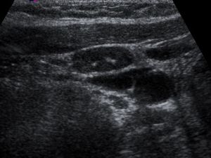
Fig. 1:
Gray-scale sonogram of patients with unpalpable metastatic nodes. Image of...

Fig. 2:
Gray-scale sonogram of patients with unpalpable metastatic nodes. Image of...

Fig. 3:
Gray-scale sonograms of patient with unpalpable metastatic nodes. Image of...

Fig. 4:
Gray-scale sonogram of patients with unpalpable metastatic nodes. Image of...

Fig. 5:
Color Doppler sonogram of a patient with metastatic nodes. Image of 64-year-old...

Fig. 6:
Gray-scale sonogram of patients with unpalpable metastatic nodes. Image of...