Primary vascular tumors of the liver in adult patients include hemangioma, epithelioid hemangioendothelioma,
angiosarcoma and hemangiopericytoma.
Some of these are benign and common,
like angioma,
others are rare and low-grade malignant tumors,
like epithelioid hemangioendothelioma.
Angiosarcoma and hemangipericytoma are very uncommon and agressive lesions.
The hepatic epithelioid hemangioendothelioma (HEH) is developing from the vascular elements of mesenchymal tissue.
Liver involvement occurs most often as a primary tumour and has a behaviour between benign hemangioma and aggressive hemangiosarcoma with extra hepatic spread.
ETIOLOGY:
The exact causes have not been clearly identified.
PATHOLOGY:
Macroscopically,
two types was described: the nodular type,
with multiple
lesions located in periphery of the liver ("peripheral pattern") and the later
confluent type with extensive lesions ("diffuse pattern").
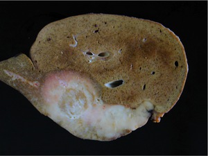
Fig. 1: Epithelioid hemangioendothelioma.The macroscopic view of a large tumor show a white mass with irregular margins and central fibrosis, findings that correspond to the imaging appearance of the intraparenchymal lesion.Hepatocytes are obliterated and replaced with a myxoid and hyalinized stroma with a progressive sclerosis and eventual calcification.
Microscopically, HEH originates from endothelial cells with positive
immunohistochemistry for factor VIII-related antigen and for the endothelial
markers CD31 and CD34.Weibel-Palade bodies are seen at electron microscopy inside tumorous cells.
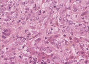
Fig. 2: Epithelioid hemangioendothelioma. Hematoxylin-eosin strain shows malignant cells in the hepatic sinusoids. Intravascular growth of these cells is responsible for the tumor infarction and central fibrosis.
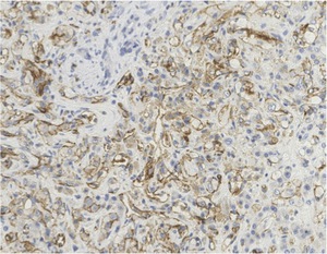
Fig. 3: Epithelioid hemangioendothelioma. Hemangioendothelioma cells are typically positive for CD31.
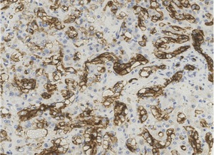
Fig. 4: Epithelioid hemangioendothelioma. Hemangioendothelioma cells are positive for CD34.
CLINICAL RELEVANCE:
Symptoms are nonspecific abdominal pain,
weight loss,
fatigue.
Other symptoms include jaundice,
fever,
hepatomegaly,
ascites or hemoperitoneum.
Budd Chiari syndrome can occur if the tumor invades the hepatic veins.
TREATMENT:
Treatment options vary from simple surveillance to surgical resection
and orthotopic liver transplant.
