This poster is published under an
open license. Please read the
disclaimer for further details.
Keywords:
Interventional non-vascular, Musculoskeletal spine, Fluoroscopy, CT, Vertebroplasty
Authors:
M. Tsitskari, D. K. Filippiadis, G. Velonakis, L. Reppas, E. Brountzos, N. L. Kelekis, A. D. Kelekis; Athens/GR
DOI:
10.1594/ecr2015/B-0339
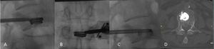
Methods and materials
During the last 42 months,
we retrospectively compared 2 groups (15 patients each) suffering from painful extreme vertebral fracture treated with standard or augmented vertebroplasty.
Group A underwent standard vertebroplasty.
Group B underwent augmented vertebroplasty with implantation of a biocompatible peek cage (KIVA implant).
Fig. 1: A: Lateral fluoroscopy view - through the working canula a nitinol coil is deployed inside the vertebral body
B: A-P fluoroscopy view - nitinol coil is deployed in the center of the vertebral body
C: Lateral fluoroscopy view - over the nitinol wire the PEEK cage is deployed; post removal of the wire the cage is filled with PMMA
D: Computed Tomography axial scan the next morning verifying proper cage positioning and no cement leakages
Standard x rays and CT scans were performed during follow-up.
Pain prior,
the morning after and at the last follow-up (average follow-up 12 months) were compared by means of a numeric visual scale (NVS) questionnaire.
Cement or implant migration were recorded.
Statistical analysis was performed with Chi-Square Tests,
Related Samples Wilcoxon Signed Rank Tests and Tests of Within Subjects Effects.