
Table 1: Alphabetical sorting and matching the underlying disease of different abdominal X-ray signs.
“A”
Definition: it is a radiologic manifestation of a focal stricture in the bowel,
on contrast studies.
Underlying Diseases:
-carcinoma of the colon (stenosing annular colorectal carcinoma) – the most frequent cause (1,2)
-lymphoma
-Crohn's disease
- chronic ulcerative colitis,
- ischaemic colitis,
- Chlamydia infection,
- tuberculosis,
- Helminthoma,
- Amoebiasis,
- Cytomegalovirus,
- villous adenoma,
- radiosurgery (2).
Radiographic features:
- The stricture shows shouldered margins and resembles an apple bite (1);
- The characteristics that suggest a colon carcinoma stricture: stricture usually has 3 to 6 cm,
mucosal irregularities and an eccentric lumen with overhanging shoulders (2);
- The characteristics that suggest a Crohn's stricture (present in up to 25% of the cases of Crohn's colitis): smooth lumen,
tapered ends that fuse into the normal bowel (2);
- The characteristics that suggest ulcerative colitis stricture are more frequent in the sigmoid colon (2);

Fig. 1: Apple core sign - contrast studies showing the appearance of an annular constricting carcinoma of the colon, from circumferential involvement of the lumen.
“B”
Definition: it is a radiologic manifestation of an irregularly marginated tapering of the inferior oesophagus.
Underlying Disease:
- The primary achalasia: is characterized by a total loss of peristalsis and the lower oesophageal sphincter relaxes poorly (4);
- The pseudoachalasia: as a result of gastric tumour involvement of the gastro-oesophageal junction (3).
Radiographic features:
- The typical smooth tapering of the gastro-oesophageal junction is compared with a bird’s beak(4);
- The oesophageal peristalsis is abnormal and the passage of the contrast material is delayed (4);
- In later stages the oesophagus is dilated and becomes tortuous,
assuming a sigmoid shape.
A large diverticula can appear over time (4).

Fig. 2: Bird’s Beak sign - contrast studies showing appearance of achalasia like a smooth tapering of the gastro-oesophageal junction.
Definition: it is a radiologic manifestation of a tapered barium column and appears when the sigmoid colon tortes on the sigmoid mesocolon(5).
Underlying Disease:
- Sigmoid volvulus (5).
Definition: it is a radiologic manifestation of a cup-shaped filling defect seen on double contrast studies.
Underlying Diseases:
-Colonic polyps;
-Colonic diverticulum (6)
Radiographic features:
- The bowler hat sign alone is a nonspecific finding for the detection of polyps (7);
-The features that suggests a colonic polyp: the bowler hat points toward the center of the long axis of the bowel(6).
The barium is trapped between the edge of the polyp and the intestinal lumen as the stalk of the polyp pulls the polyp against the adjacent wall (8).
- The features that suggest a colonic diverticulum: the bowler hat points away from the center of the long axis of the bowel (6).

Fig. 3: Bowler Hat Sign - double contrast study showing cup-shaped filling defects that point away from the lumen, representing colonic diverticulum.
“C”
Definition: it is a radiologic manifestation created by a large flat ulcer with heaped-up edges.
Underlying disease:
- Malignant gastric ulcer
Radiographic features:
- The edges of the ulcer trap a lenticular barium collection that is convex relative to the lumen,
when the edges are folded upon themselves during compression (9).
Curiosity…
Russell Daniel Carman (1875–1926) was a professor of Roentgenology at the medical schools of St Louis University and Washington University.
His work pioneered gastrointestinal radiology,
resulting in the publication of The Roentgen Diagnosis of Diseases of the Alimentary Canal,
in 1917.
In 1925 he became ill and his colleagues performed a fluoroscopic examination of him.
When he saw the films he said: “cancer of the stomach,
inoperable”(9).
Definition: it is a radiologic manifestation of the raised right hemidiaphragm delineated by subdiaphragmatic air,
representing bowel gas interposed between the liver and the hemidiaphragm (10,11).
Underlying disease:
-Pseudopneumoperitoneum
Radiographic features:
- An haustral pattern overlapping the upper border of the liver shadow,
like a crescent-shaped (subdiaphragmatic radiolucency) (10);
- Between the right hemidiaphragm and the liver stays the lumen of the transverse colon filled with gas (10);
- It occurs in 0.25% to 0.28% of the general population with a slight increase in individuals aged above 60 years (12);
- It is more frequent in men than in women (4:1 ratio) (12);
- This sign can be related with the Chilaiditi’s syndrome (symptomatic hepatodiaphragmatic colonic interposition) (11);
- A computed tomography scan is necessary to establish to diagnosis,
when a radiography cannot clearly determine whether the subdiaphragmatic air is free or intraluminal(12).
Curiosity...
Demetrious Chilaiditi was the author who described in first hand this sign in the year of 1910 (10).

Fig. 4: Chilaiditi’s Sign: plain film showing interposition of bowel gas between the liver and the right hemidiaphragm.
Definition: it is a characteristic abdominal radiographic sign of sigmoid volvulus and consists of a greatly distended,
air-filled loop of sigmoid colon extending from the pelvis (1).
Underlying diseases:
- Sigmoid volvulus;
- Closed loop small bowel obstructions (13)
Radiographic features:
- A oblique line that is formed by the apposition of the medial walls of the dilated bowel,
resembles the cleft of a coffee bean (1);
- The “coffee bean” can appear from the pelvis and its apex can be at T10 level to the left or right of midline (1).

Fig. 5: Coffee-bean Sign - plain film showing dilated sigmoid colon in sigmoid volvulus.
Definition: it is a radiologic manifestation wich associates an abrupt termination of gas pattern,
within the proximal colon at the level of the radiographic splenic flexure,
and a decompression of the distal colon (14).
Underlying diseases:
-Acute pancreatitis;
-Carcinoma of colon;
-Inflammatory bowel disease;
-Mesenteric ischaemia.
Features:
- In acute pancreatitis the inflammatory exudate extends into the phrenicocolic ligament by spreading through the lateral attachment of the transverse mesocolon (14);
- The functional spasm and/or mechanical narrowing of the splenic flexure,
at the level where the colon returns to the retroperitoneum,
are caused by infiltration of the phrenicocolic ligament (14);
- Though originally described in abdominal radiographs,
this sign has also been demonstrated in contrast enemas and computed tomography (14).
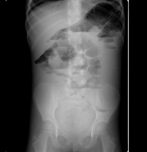
Fig. 6: Colon Cutoff Sign: abdominal radiography showing dilated transverse colon to splenic flexure, in this case was associated with pancreatitis.
Definition: it is a radiologic manifestation of constricted and twisted lumen usually seen in motility disorder of the oesophagus.
Underlying disease:
- Diffuse oesophageal spasm
Radiographic features:
- Contrast studies show simultaneous and uncoordinated contraction,
that obliterate the lumen (15);

Fig. 7: Corkscrew Oesophagus: contrast study showing constricted and twisted lumen in diffuse oesophageal spasm from abnormal tertiary contractions.
Definition: it is a radiologic manifestation of air beneath the diaphragm (16).
Underlying disease:
- Pneumoperitoneum
Differential diagnosis (16):
- Chilaiditi syndrome;
- Subdiaphragmatic abscess;
- Omental fat interpositioned between the liver and diaphragm;
- Subpulmonary pneumoperitoneum;
- Enlarged gastric bubble.
Radiographic Features:
-The radiography of the abdomen in the erect position is the best mean of detecting pneumoperitoneum (17).
-Subdiaphragmatic free air is the presence of free extraluminal air,
in the anterior subhepatic space (16,18).
-The abdominal radiography can detect the amount of 1 ml of free air,
but the patient needs to stay in the upright position about 10 minutes,
for the air to rise (16,18).

Fig. 8: a) Crescent Sign: plain film showing appearance of a sliver of air usually beneath the both hemidiaphragms in pneumoperitoneum. b) Chilaiditi’s Sign: plain film showing interposition of bowel gas between the liver and the right hemidiaphragm.

Fig. 9: Crescent Sign: abdominal radiography showing air beneath both hemidiaphragms, in relation with pneumoperitoneum.
Definition: it is a radiologic manifestation of free air under the central diafragmatic tendon and appears like an arcuate lucency superimposed on lower thoracic spine that projects caudad to the heart (19).
Underlying disease:
- Pneumoperitoneum
Radiographic features:
-The “Cupola” indicates the inverted cup-shaped configuration of the radiolucency (19);
-The superior border of the radiographic radiolucency is well defined and the inferior margin is not very well delineated (19);
-The cupola sign represents free intraperitoneal air within the median subphrenic space which may lead further investigations (19).

Fig. 10: Cupola sign: abdominal radiography showing free intraperitoneal air under the central diaphragmatic tendon.
“D”
Definition: it is a radiologic manifestation represented as a dilatation of the proximal stomach and duodenum,
with little or no air distally,
seen in newborns and infants (20).
Underlying disease (20):
-Congenital obstruction (duodenal web;duodenal atresia;duodenal stenosis;annular pancreas);
-Midgut volvulus;
-External compression of the dudenum (preduodenal portal vein; annular pancreas; midgut volvulus; Ladd bands).
Radiographic features:
-The bubble on the left side of the midline filled with air represents the stomach and the bubble on the right of the midline represents the proximal duodenum(20);
-This sign is reproducible with upper gastrointestinal studies and sonography(20);
-The appearance,
prominence and presence of air distal to the obstruction are determinate by the specific duodenal structural anomaly causing the obstruction(20);
-If the cause of the obstruction is the duodenal atresia,
it can produces complete obstruction,
and there is no distal bowel gas(20);
- Duodenal web and duodenal stenosis are partially obstructing anomalies that allow passage of some air distal to the obstruction and resulting in a less prominent double bubble(20).

Fig. 11: Double Bubble Sign: abdominal radiography showing two air-filled structures in the upper abdomen, with no air distally. The proximal bubble in the left side filled with air represents the stomach and the second bubble to the right of the midline represents the proximal duodenum.

Fig. 12: Double Bubble Sign: oral contrast study showing two contrast-filled structures in the upper abdomen, with no contrast distally. The proximal structure in the left side filled with contrast represents the stomach and the second structure to the right of the midline represents the proximal duodenum.
Definition: it is an upper gastrointestinal sign represented in the gastrointestinal series where barium is separated into two columns within an abnormal pyloric channel.
Underlying disease (21,22):
- Hypertrophic pyloric stenosis.
Radiographic features:
-The elongated pylorus in the contrast studies is represented by two parallel lines of barium from the pre-pyloric region to the base of the cap,
interposed by a radiolucent band (21);

Fig. 13: “Double Track Sign” - Contrast study showing redundant mucosa separating barium into two columns in pyloric channel.
“F”
Definition: it is a radiologic manifestation of the usually invisible falciform ligament,
which becomes visible when is surrounded by intraperitoneal air.
Underlying disease:
- Pneumoperitoneum
Radiographic features:
-The falciform ligament connects the anterior abdominal wall to the anterosuperior surface of the liver(23);
-Under normal circumstances the falciform ligament is invisible(23);
-The falciform ligament become visible,
as a vertical band of soft tissue parallel to the right border of the spine,
if it is surrounded by intraperitoneal free air(23);
-This sign also appears in computed tomography scan images(23).

Fig. 14: Falciform Ligament Sign - abdominal radiography showing the falciform ligament from surrounding air, in pneumoperitoneum.
Definition: it is a radiologic manifestation of transient transverse oesophageal folds on double contrast oesophagram.
Underlying diseases (24):
-Gastro-oesophageal reflux (GER);
-Eosinophilic oesophagitis.
Radiographic features:
-In a “feline oesophagus”,
the oesophagus presents with fine,
transverse folds crossing the entire lumen(24);
- It is called “feline” because it mimics the appearance of the folds in the distal oesophagus of the cat (24);
-The folds are linear/angular,
they have 1-2mm wide,
extend circumferentially around the lumen and are present in different parts of the oesophagus (24);
-The folds appear trasiently during upright double-contrast oesophagography and barium swallowing (24);
Definition: it is a radiologic manifestation on supine abdominal position of an oval radiolucency with the shape of an American football (25).
Underlying diseases:
-Large pneumoperitoneum in infants (necrotising enterocolitis; peptic ulcer disease);
-Bowel obstruction with secondary perforation;
-Endoscopic perforation.
Radiographic features:
-The long axis of the “football” ball has a cephalo-caudad orientation (25),
- The blunted ends are delineated by the diaphragm and pelvic floor (25);
- The full-blown football sign could not be present when small amounts of free gas are present (26);
-This sign is more common in infants (25).
-It is present in 2% of adults(25).
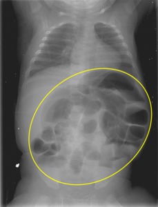
Fig. 15: Football Sign - abdominal radiography showing a large oval radiolucency demarcated by the parietal peritoneum of the abdominal wall.
“H”
Definition: it is a radiologic manifestation that appears as a thin line in profile at the neck of gastric ulcer,
representing the thin rim of undermined gastric mucosa (27).
Underlying disease:
-Benign gastric ulcer
Radiographic features:
- The Hampton's Line is like a thin straight line,
seen in profile,
across the neck of a gastric ulcer,
on contrast studies (27).
-The line shows the benign nature of the gastric ulcer once the resistant mucosa overhangs the more rapidly destroyed submucosa(27).
Curiosity…
Aubrey Otis Hampton,
who was a worker at Massachusetts General Hospital,
described for the first time the Hampton line sign,
Hampton hump and Hampton maneuver (28).
“L”
Definition: it is a radiographic pattern of a rigid and non-haustral colon (29).
Underlying disease:
-Chronic ulcerative colitis
Radiographic features:
-When it is present there is a complete loss of haustral markings in the diseased section of colon (29);
“M”
Definition: it is the radiographic appearence of a triradiate radiolucent shadow seen in the right upper quadrant of the abdomen (30).
Underlying disease:
-Gas fissuring within gallstones
Radiographic features:
- It is a rare sign (30);
- The gas in the interior of the calculus or Mercedes-Benz sign is caused by the development of fissures in the process of crystallization (31);
-It can be confused for gas and feces in a superimposed loop of bowel (30);
- The typical triradiate pattern is seen if the x-ray beam aligns with the longitudinal axis of the calculus (30)
“P”
Definition: it represents an apparent visualization of both sides of bowel wall when two dilated loops of bowel abut each other (32).
Underlying disease:
-Apparent visualization of both sides of bowel wall
Differential diagnosis (33):
- Free fluid in the peritoneal cavity;
- Adjacent loops of bowel with thickened walls.
Radiographic features:
-The appearance of abnormal separation of the walls of adjacent bowel loops can be produced by intraluminal fluid (33);

Fig. 16: Pseudo-Rigler’s Sign - abdominal radiography showing both sides of bowel wall with dilated loops of bowel abut each other.
Definition: it is a radiographic manifestation of an outpouching created on lesser curve by distorted muscle in hypertrophic pyloric stenosis (34,35).
Underlying disease:
-Hypertrophic pyloric stenosis
Radiographic features:
-The pyloric tit sign represents contrast material trapped between the peristaltic wave and the muscle (35).
The contrast material is compressed between a peristaltic wave and the impression of the pyloric muscle upon the adjacent portion of the stomach (34).

Fig. 17: Pyloric Tit Sign – contrast study showing an outpouching created on lesser curve by distorted muscle in hypertrophic pyloric stenosis.
“R”
Definition: appearance in “reversed 3” of inner margin of duodenal loop from enlarged head of pancreas,
with tethering at ampulla (36).
Underlying diseases:
-Pancreatitis;
-Duodenal adenocarcinoma;
-Pancreatic adenocarcinoma.
Definition: it is a radiographic manifestation of gas on both sides of the bowel wall (37).
Underlying disease:
-Pneumoperitoneum
Radiographic features:
-It can be called the double wall sign (37);
-If intraperitoneal free gas is present in a moderate amount,
it will be accumulated between bowel loops,
permitting the visualization of the outer walls of the bowel (37);
Curiosity …
Rigler (1896-1979) described for the first time the sign of pneumoperitoneum that now bears his name.
He established the first postgraduate course in radiology and he built a legacy of radiologic teaching,
instituting the interdepartmental radiology conference at the University of Minnesota (37).

Fig. 18: Rigler sign - abdominal radiography showing free air outlining the small bowel wall, indicating pneumoperitoneum.
“S”
Definition: this sign shows dilated loops of the small bowel,
on a radiographic plain film.
Underlying disease:
-Focal area of adynamic ileus of an isolated small-bowel segment (14).
Radiographic features:
- It is the best known of all the abdominal signs (14);
-The sentinel loop sign is a nonspecific finding at the radiography (38).

Fig. 19: Sentinel Loop Sign - abdominal radiography showing dilated loops of small bowel, in a patient with an acute pancreatitis.
Definition: it is a radiographic manifestation of the dilatation of multiple small bowel loops in the left upper quadrant.
Underlying disease:
-Mechanical small bowel obstruction
Radiographic features:
-The stepladder formation of the dilated small intestinal coils across the abdomen is typical of a partial or total obstructive lesion (39);
-The demonstration of Kerkring folds are pathognomonic of the small bowel,but they are not always observed (40).
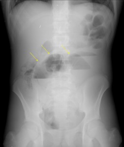
Fig. 20: Stepladder Appearance - abdominal radiography showing dilated loops of the small bowel in the left upper quadrant in a mechanical small bowel obstruction.
Definition: it is a radiographic manifestation of multiple,
obliquely or horizontally orientated small pockets of air in a fluid dilated small bowel loop (41).
Underlying disease:
-Mechanical small bowel obstruction (SBO)
Radiographic features:
- This sign can also be called the string of pearls sign,
and it can appear in upright or in decubitus on abdominal radiographs (41);
- The sign first appears on horizontal-beam radiographs and then on supine radiographs (41);
-The ovoid or rounded appearance of the trapped air are produced by the meniscal effect of the surrounding fluid (41);
-The visual aspect of the string of pearls sign depends if air,
fluid-filled bowel loops or peristaltic hyperactivity are present (41);
-It's knowledge avoid the delay of the diagnosis of SBO when the clinical manifestations are confusing (41).

Fig. 21: String of Beads Sign - abdominal radiography showing linearly arranged small pockets of air in a fluid-dilated small bowel loop.
Definition: it is a contrast study manifestation of a very thin luminal contrast seen in terminal ileum from spasm and eventually from fibrosis (42).
Underlying diseases:
-Crohn disease;
-Carcinoid tumour.
Radiographic features:
-It is observed most frequently in the terminal ileum (42);
-The gastrointestinal string sign results from a severe narrowing of a bowel loop,
caused by spasm or stenosis,
associated with severe ulceration,
which makes the lumen resemble a frayed cotton string (42).

Fig. 22: String Sign – contrast study showing a string-like appearance of a contrast filled bowel loop caused by severe narrowing of a bowel loop.
“T”
Definition: it is like an indentation in barium contrast or in gas filled bowel lumen caused by submucosal infiltration and resembles a thumbprint (43).
Underlying diseases:
-Inflammatory bowel disease;
-Infection (pseudomembranous colitis);
-Diverticulitis;
-Mucosal/submucosal haemorrhage.
Radiographic features:
-The pathophysiologic mechanism is based on the submucosal oedema and on hemorrhage from capillary leakage (43).

Fig. 23: Thumbprinting – abdominal radiography showing ‘thumbprinting’(arrows). The normal haustral folds are replaced by wide transverse thickened bands.
"U"
Definition: it is a contrast study manifestation of organoaxial rotation of the stomach in gastric volvulus resulting in reversed positions of lesser and greater curvatures (44,45).
Underlying disease:
-Gastric volvulus
Radiographic features:
-This sign is related with 180 degrees anterior organoaxial torsion(44),
the stomach rotates around its long axis (45);
-It occurs when the greater curvature of the stomach lies above the lesser curvature and the posterior wall faces forward,
in an upside-down shape (44);
-The rotation commonly occurs from right to left (45);
-The pylorus rotates anteriorly and superiorly and when this occurs,
the distal gastric segment and the cardioesophageal junction usually become obstructed (45).

Fig. 24: Upside Down Stomach – contrast study showing an intratoracic stomach with reversed positions of lesser and greater curvatures, representing a gastric organoaxial volvulus.


 Crescent Sign: plain film showing appearance of a sliver of air usually beneath the both hemidiaphragms in pneumoperitoneum. b) Chilaiditi’s Sign: plain film showing interposition of bowel gas between the liver and the right hemidiaphragm.")


. The normal haustral folds are replaced by wide transverse thickened bands.")
